The oculomotor nerve is the third cranial nerve.
Function
Together with the trochlear nerve and the abducens nerve, the oculomotor nerve is responsible for eye movements.
Nuclei
The oculomotor nerve originates from two nuclear complexes. The oculomotor nucleus is a somatomotor nucleus responsible for movements of the striated extraocular muscles. The accessory oculomotor nucleus is a visceromotor nucleus responsible for movements of the smooth intraocular muscles1.
Anatomical course
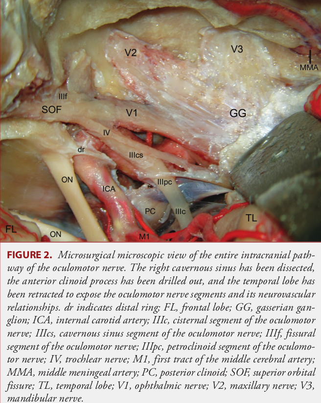
Various classifications of the oculomotor nerve have been described in the literature for anatomical delineation. The classification described here is based on the publication by Iaconetta et al.2 and divides the oculomotor nerve into five anatomical segments.
Cisternal segment
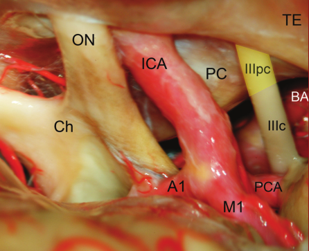
In the cisternal segment, the oculomotor nerve courses within the interpeduncular cistern. The oculomotor nerve exits the midbrain medial to the cerebral peduncles in the lateral portion of the interpeduncular fossa and courses anterolaterally and superiorly to the posterior margin of the petroclinoid dural duplication, which forms the anterior end of this segment. Here, the oculomotor nerve courses in close relation to the PCA and the SCA, as well as their perforators.
Petroclinoid segment
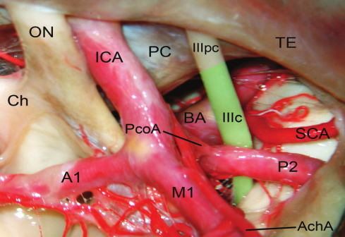
After the cisternal segment, the oculomotor nerve continues into the petroclinoid segment, which extends to its entry into the lateral wall of the cavernous sinus. The floor of the petroclinoid segment is formed by the oculomotor triangle.
Compression of the oculomotor nerve
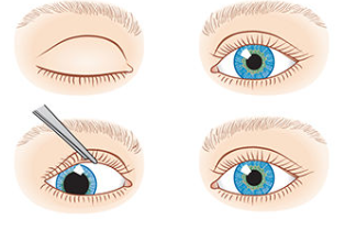
Compression of the oculomotor nerve typically initially manifests as a dilated pupil (5-6 mm)3. Compression may be caused by uncal herniation, a progressive aneurysm of the posterior communicating artery, or aneurysmal dilatation at the basilar artery bifurcation. A disturbance of ocular motility occurs in most patients after sustained compression for more than 24 hours4.
Illustration of deficit symptoms
References
Trepel, Martin, and Erich Mayer-Fally. "Neuroanatomie." Struktur und Funktion 18.7 (2017): 30021-7. ↩ ↩
Iaconetta, Giorgio, et al. "The oculomotor nerve: microanatomical and endoscopic study." Neurosurgery 66.3 (2010): 593-601. ↩ ↩
Handbook of Neurosurgery. Greenberg M, ed. 9th Edition. New York: Thieme; 2019. ↩ ↩ ↩ ↩