Olfactory groove meningiomas are Meningiomas arising from the Dura mater or the Arachnoid mater of the olfactory groove and may occur as WHO Grade 1–3 tumors depending on their histological type.
Epidemiology
Olfactory groove meningiomas are rare and, together with Planum sphenoidale meningiomas, account for approximately 2 % of all primary intracranial tumors[^1]. Olfactory groove meningiomas occur somewhat more frequently in women than in men, with a ratio of approximately 1.45:1[^9].
Symptoms
Olfactory groove meningiomas often remain asymptomatic for a relatively long period and are then frequently diagnosed once they have already reached a considerable size. Unilateral or bilateral loss of the sense of smell is usually one of the first symptoms, although patients often do not notice it. Most patients initially present with headaches, visual impairment, or cognitive changes[^9].
Vascular supply
Olfactory groove meningiomas frequently receive their vascular supply via the Anterior ethmoidal artery and the Posterior ethmoidal artery, as well as via the anterior branches of the Middle meningeal artery and meningeal branches of the Ophthalmic artery[^2]. In larger olfactory groove meningiomas, the vascular supply may also arise from direct branches of the Anterior cerebral artery or the Anterior communicating artery[^2].
Imaging
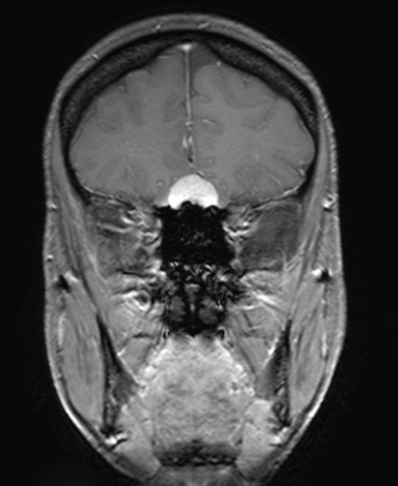
The gold standard for imaging olfactory groove meningiomas is MRI imaging. digital subtraction angiography is generally not necessary for preoperative planning, as the tumor’s vascular relationships can usually be adequately visualized on conventional MRI[^3].


Surgical approaches
Olfactory groove meningiomas can be reached and resected through various surgical approaches.
Subfrontal approach
The subfrontal approach offers the advantage of early devascularization of the tumor at the Skull base. In addition, this approach allows vascularized pericranium to be harvested for skull-base reconstruction. A disadvantage of this approach is opening the Frontal sinus, with the associated increased risk of a Cerebrospinal fluid fistula and infection. This approach also involves transection of the anterior portion of the Superior sagittal sinus[^3].
Pterional approach
Olfactory groove meningiomas can also be reached through a pterional craniotomy. The advantage of this approach is early visualization of the optic nerve and the internal carotid artery before tumor manipulation. The basal cisterns are also reached early in the procedure, allowing cerebrospinal fluid to be drained for improved surgical visualization. A disadvantage of this approach is the limited angle of access to the superior portion of the tumor, which often requires retraction of the frontal lobe. Furthermore, skull-base reconstruction is difficult through this approach[^3] [^4].
Supraorbital keyhole approach
Another option is a supraorbital approach through a keyhole. One study demonstrated a shorter hospital stay and less postoperative edema compared with conventional approaches[^7].
Interhemispheric approach
The advantage of an interhemispheric approach is direct exposure of the tumor with minimal brain retraction and without opening the frontal sinus [^8]. The disadvantage of this approach is the difficult access to the vascular supply of the olfactory groove meningioma. In addition, the operative corridor is long and narrow, which may result in contusions of the frontal lobe. Depending on the individual anatomy, Bridging veins may make this approach more difficult[^3].
Endoscopic endonasal approach
The endoscopic endonasal approach offers the advantage of direct access to the tumor with minimal brain retraction and the possibility of early tumor devascularization[^5]. However, this approach has a steep learning curve and, depending on the extent of tumor spread, may not provide access to all intracranial portions of the tumor[^3] [^6].