The frontal sinus is a paired, mucosa-lined cavity in the Frontal bone and is one of the paranasal sinuses.
Development
The frontal sinus begins to develop from the second year of life and is radiologically visible from the age of 8.
Anatomy
The frontal sinus is lined with respiratory epithelium. Medially, it drains via the Frontonasal duct, and laterally, the frontal sinus drains into the Middle nasal meatus. In adults, the frontal sinus extends an average of 2.8 cm superiorly from the Nasion and is generally located no more than 5 mm lateral to the Midpupillary line[^2].
Imaging

Clinical relevance
Frontal sinus fractures
Fractures of the posterior wall of the frontal sinus may lead to a delayed intracranial Abscess, Cerebrospinal fluid fistula with subsequent Meningitis, or a Mucocele[^1].
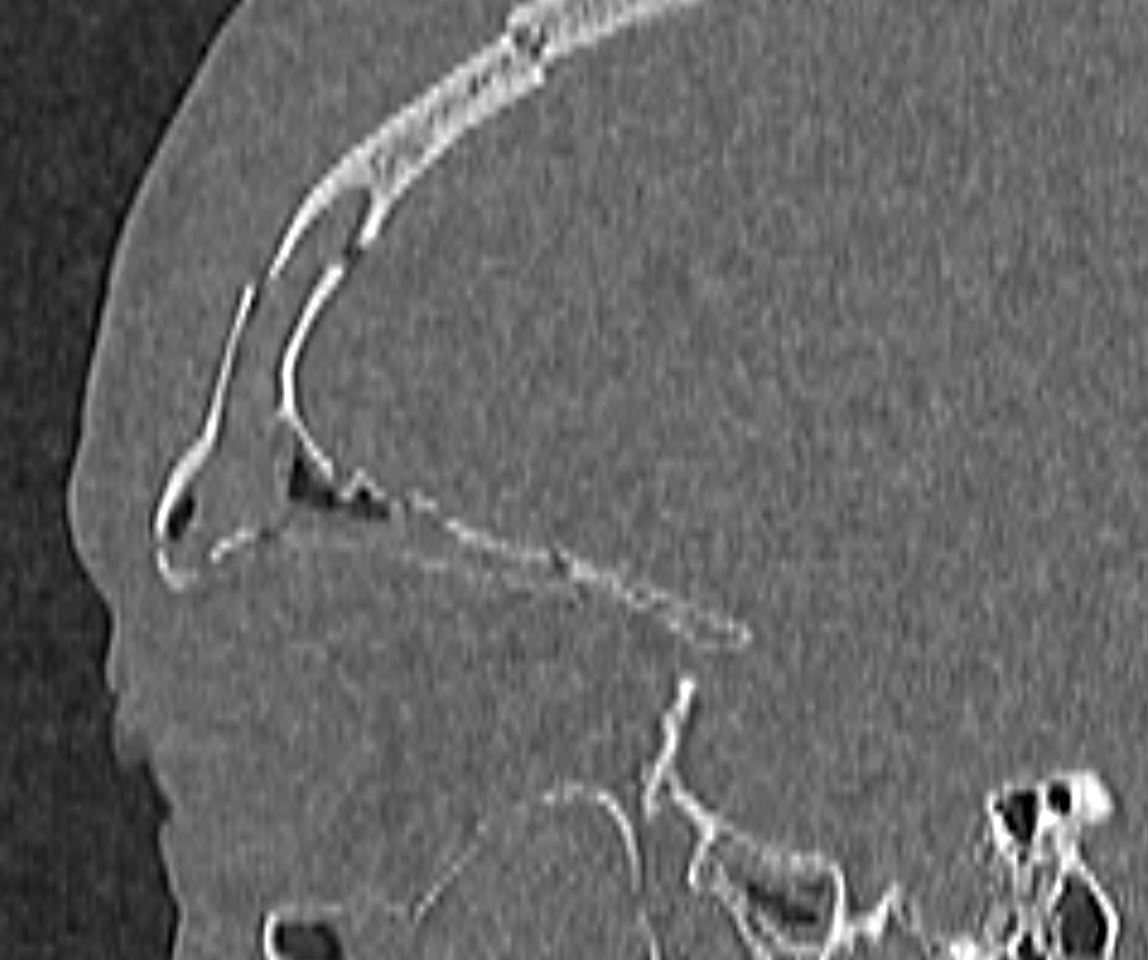
Cranialization of the frontal sinus
During cranialization of the frontal sinus, the frontal sinus mucosa should be removed and the frontal sinus should be obliterated with, for example, adipose tissue or muscle[^4]. To create an additional protective layer, a pedicled flap of Galea aponeurotica may be used, for example[^3].