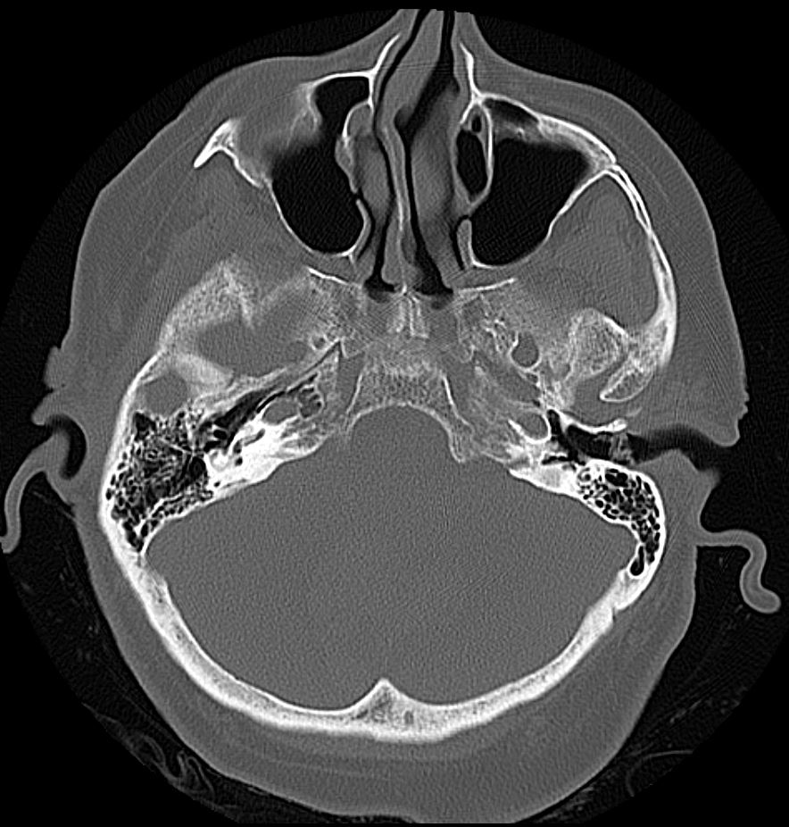
The occipital bone is a cranial bone and part of the neurocranium.
Anatomy
The occipital bone borders the two Lambdoid suture cranially via the Parietal bones, the Temporal bone laterally, and the Sphenoid bone inferiorly.
The occipital bone consists of several anatomical parts that together form a complex structure[^1]:
Basilar part
The basilar part is located ventral to the Foramen magnum and adjacent to the petrous part of the temporal bone. Ventrally, the basilar part fuses with the sphenoid bone during puberty (tribasilar bone), thereby forming the Clivus. Both the superior pharyngeal constrictor muscle and the pharyngeal raphe attach to the pharyngeal tubercle, which is located on the inferior surface of the basilar part. The surface of the basilar part is smooth, and its internal surface contains the clival region, which plays a central role in skull base surgery.
Lateral part
The bilateral lateral parts, also called the condylar parts, are located lateral to the foramen magnum. Both sides have a kidney-shaped process, the Occipital condyle, which articulates with the first cervical vertebra via the atlanto-occipital joint. The Condylar canal lies dorsally and is traversed by the condylar emissary vein, which connects the external vertebral venous plexus with the Sigmoid sinus. The Hypoglossal nerve passes through the Hypoglossal canal, which runs through the lateral part of the occipital bone.
Squamous part of the occipital bone
The squamous part is the largest part of the occipital bone. A palpable bony prominence along the midline of the external surface of the bone, the External occipital protuberance, serves as the site of origin of the trapezius muscle. In addition, the external surface has three arched bony ridges, known as the nuchal lines:
The Linea nuchae suprema runs above the external occipital protuberance and serves as the attachment site for the epicranius muscle and the Galea aponeurotica.
The Linea nuchae superior runs slightly below it. It serves as the attachment site for the trapezius muscle, Musculus sternocleidomastoideus, and splenius capitis muscle.
The Linea nuchae inferior runs slightly lower, and the semispinalis capitis muscle attaches above it.
On its internal surface, the squamous part of the occipital bone has depressions formed by intradural venous channels: the Sinus sagittalis superior, the Sinus transversi, and the Sinus sigmoideus. Above the Sulcus sinus transversus is a depression for the brain’s Lobus occipitalis, and the Cerebellum lies in the fossa below it.
Figure