Optic chiasm

Last edit by Alaric Steinmetz on

Synonyms: Optic chiasm

ICD-11: XA5TX3

The optic chiasm is the site where the two optic nerves cross.

Anatomy

The optic chiasm is located in the middle cranial fossa within the sulcus chiasmatis of the sphenoid bone. In its immediate vicinity, the floor and anterior wall of the third cerebral ventricle meet. Inferior to the chiasm lies the sella turcica with the pituitary gland, and posteriorly lies the pituitary stalk.

Anatomical variants

Variant Optic chiasm location1 Frequency2
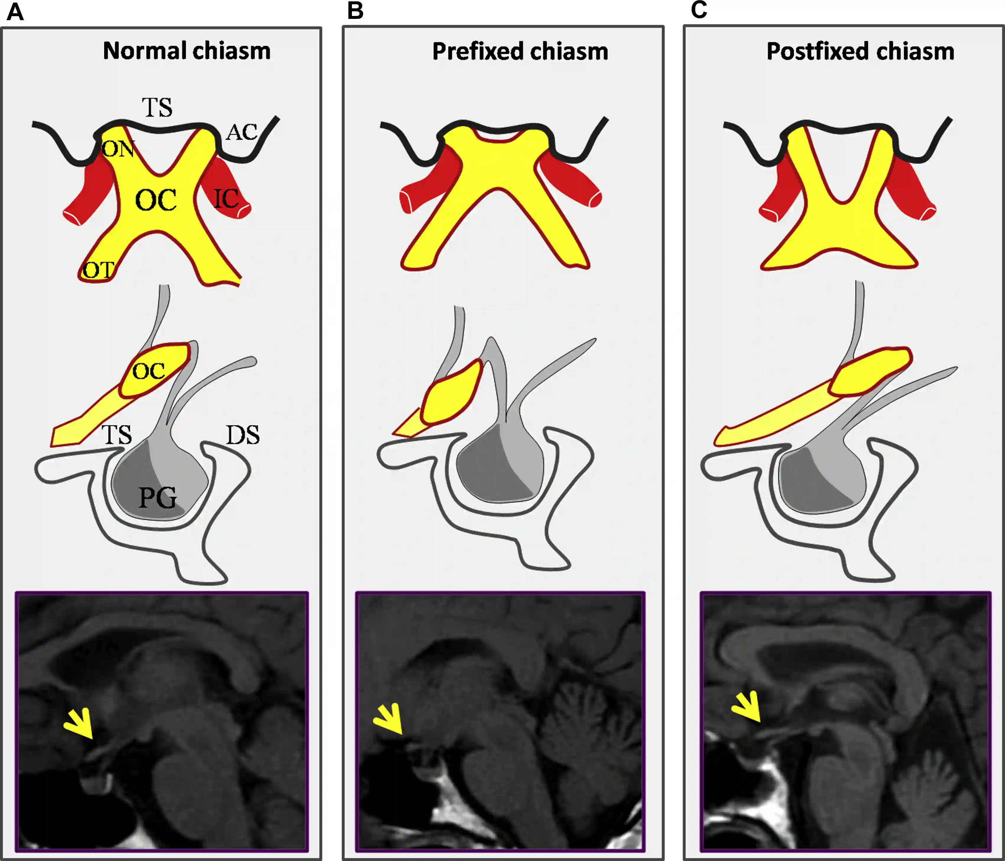
Normal Above the diaphragma sellae ~ 80 %
Prefixed Above the tuberculum sellae ~ 10 %
Postfixed Above the dorsum sellae ~ 10 %

Anatomical variations of the optic chiasm
Schematic and MRI illustrations of the three anatomical variations of the optic chiasm. AC, anterior clinoid; DS, dorsum sellae; IC, internal carotid artery; OC = optic chiasm; ON = optic nerve; OT = optic tract; PG = pituitary gland; TS = tuberculum sellae. Figure adapted from Prieto et al. World Neurosurgery 2015.

Clinical relevance

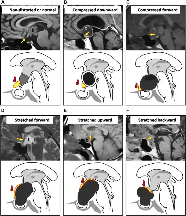
Tumors in the region of the pituitary gland, such as pituitary adenomas or craniopharyngiomas, can cause displacement or compression of the optic chiasm, resulting in visual field defects, typically a bitemporal hemianopia, or visual impairment.

Displacements of the optic chiasm
Schematic and MRI illustrations of possible displacements of the optic chiasm. Figure adapted from Prieto et al. World Neurosurgery 2015.

Imaging

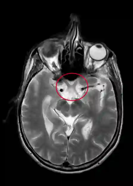
The optic chiasm can be visualized on MRI.

Optic chiasm on MRI
Visualization of the optic chiasm (within the red marking) on an axial T2 MRI sequence of the skull.

References


    Article information

    Article created by
    Last edited:
    on
    Revision history
    Show revision history (2)

    2 edits by 1 author

    Edit · Alaric Steinmetz ·

    No content change recorded

    Article created on · Alaric Steinmetz

    Author list:
    Newsletter