Chronic Subdural Hematoma
Last edit by Alaric Steinmetz on
Synonyms: pachymeningitis hemorrhagica interna, cSDH
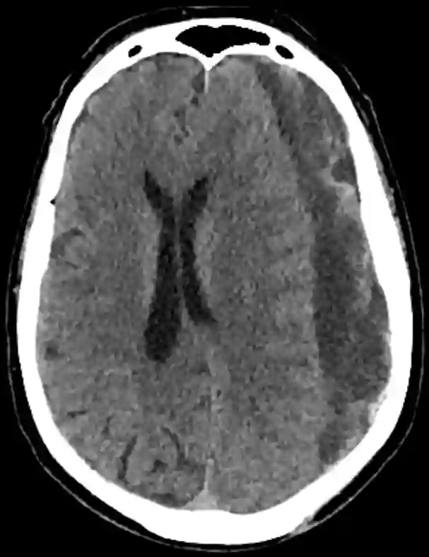
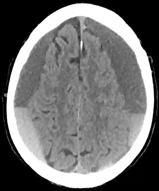
Chronic subdural hematoma is a common neurosurgical condition that occurs particularly often in older adults after a fall with head impact. It is a chronic blood collection between the brain and the dura mater.
Epidemiology
In patients over 70 years of age, chronic subdural hematoma occurs with an incidence of 58/100000 persons1.
Causes and Pathophysiology
Chronic subdural hematoma often occurs several weeks after a fall and probably develops from an initially minor acute subdural hematoma2. Blood in the subdural space triggers an inflammatory response, resulting in the migration of fibroblasts and the formation of neomembranes on the cortical and dural surfaces of the hematoma3. This then leads to the formation of neocapillaries, enzymatic fibrinolysis, and subsequent liquefaction of the blood clot4. Fibrin degradation products are redistributed into new blood clots and inhibit hemostasis. The course of a chronic subdural hematoma is determined by a balance between plasma effusion and/or recurrent bleeding originating from the neomembranes on one side and resorption of the hematoma on the other5 6.
Risk Factors
The use of medications that inhibit platelet aggregation (such as aspirin or clopidogrel) or affect coagulation promotes the development of chronic subdural hematoma. Known risk factors for the development of chronic subdural hematoma include age, alcohol dependence, epileptic seizures, cerebrospinal fluid shunts, and cerebral atrophy7 8. A history of trauma is not always present. 29-38% of all patients have no recollection of a fall9.
Symptoms
Symptoms of chronic subdural hematoma often develop slowly and frequently present with mild symptoms such as headache, confusion, or speech disturbances10. Symptoms may also be more pronounced and manifest as hemiparesis progressing to hemiplegia. Seizures of varying severity, including comatose states, may also occur11. The Markwalder classification can be used to quantify the clinical severity for academic purposes; depending on the clinical condition, it classifies patients into five different grades.
Imaging
The gold standard for the imaging diagnosis of chronic subdural hematoma is CT imaging of the head. The diagnosis can also be made with MRI.


Treatment Options
In some cases, chronic subdural hematoma is diagnosed incidentally, and the patient is asymptomatic. If the hematoma is also small in such a case, conservative treatment may be an option.
Surgical Management
Burr-Hole Trepanation and Craniotomy
Surgical removal of a chronic subdural hematoma is one of the most common neurosurgical procedures. Typically, two burr holes are created on the affected side using a trephine, through which the chronic hematoma can be irrigated. In some cases, there are extensive septations between the liquid portions of the hematoma, which significantly complicate the operation. The burr holes created in the calvarium are usually covered only with a titanium plate for cosmetic reasons, to prevent the skin from sinking in at this site. In rare cases, the hematoma components cannot be removed as intended through the burr holes, and the procedure must be extended to include a craniotomy. The procedure can be performed under general anesthesia or, if necessary, under local anesthesia. Even at an advanced age, most patients benefit greatly from the operation and recover very quickly from the procedure.
Surgical Risks
- Seizures, including refractory status epilepticus
- Intracerebral hemorrhage in approximately 0.7 - 5%12
- Pneumocephalus
- Recurrence of chronic subdural hematoma
Embolization of the Middle Meningeal Artery
In patients for whom surgical treatment with burr-hole trepanation or craniotomy is not the first-line treatment, endovascular embolization of the middle meningeal artery may be performed as treatment. Neomembranes of a chronic subdural hematoma contain neovascularization, which contributes to recurrent bleeding after surgical removal13. During embolization of the middle meningeal artery, ophthalmic collaterals should be avoided whenever possible.
Pharmacological Treatment
Because of the inflammatory pathogenesis of chronic subdural hematomas, therapies with dexamethasone have also been investigated for the treatment of chronic subdural hematomas. In a multicenter study of 748 patients, additional treatment with dexamethasone was shown to result in worse outcomes and more frequent complications, but fewer repeat operations14.
Outcome
Older patients also often benefit greatly from surgery, as the procedure may be performed under local anesthesia in high-risk patients. The mortality rate for surgical treatment of chronic subdural hematoma is approximately 0.5-8 % in the literature15 16. The 1-year mortality rate for chronic subdural hematoma is approximately 32 %17.
References
Kudo H, Kuwamura K, Izawa I, Sawa H, Tamaki N: Chronic subdural hematoma in elderly people: present status on Awaji Island and epidemiological prospect. Neurol Med Chir (Tokyo) 32:207–209, 1992 ↩ ↩
Handbook of Neurosurgery. Greenberg M, ed. 9th Edition. Thieme; 2019. ↩ ↩ ↩ ↩ ↩ ↩ ↩ ↩ ↩ ↩ ↩ ↩ ↩ ↩
Labadie EL, Sawaya R. In: Fibrinolysis in the Formation and Growth of Chronic Subdural Hematomas. Fibrinolysis and the Central Nervous System. Philadelphia: Hanley and Belfus; 1990:141-148. ↩ ↩
Drapkin AJ. Chronic Subdural Hematoma: Pathophysiological Basis of Treatment. Br J Neurosurg. 1991;5:467-473. ↩ ↩
Book: Neurosurgical Knowledge Update - A comprehensive review. Robert E. Harbaugh et al. Thieme Verlag. ↩ ↩ ↩ ↩ ↩ ↩
Ogasawara K, Koshu K, Yoshimoto T, et al. Transient Hyperemia Immediately After Rapid Decompression of Chronic Subdural Hematoma. Neurosurgery. 1999;45:484-489. ↩ ↩ ↩ ↩
Hutchinson, Peter J., et al. "Trial of Dexamethasone for Chronic Subdural Hematoma." New England Journal of Medicine (2020). ↩ ↩
Sambasivan M. An Overview of Chronic Subdural Hematoma: Experience with 2300 Cases. Surg Neurol. 1997;47:418-422. ↩ ↩