Sphenoid wing meningiomas are Meningiomas that arise from the Dura mater of the Os sphenoidale.
Epidemiology
Sphenoid wing meningiomas are the third most common meningiomas and account for approximately 15-20 % of all intracranial meningiomas[^2]. These tumors most commonly occur in the fourth decade of life, and as with meningiomas in other locations, women are more frequently affected[^3].
Symptoms
The most common symptoms leading to the initial diagnosis are headache, seizures, and visual disturbances. The en plaque hyperostosing forms occur almost exclusively in women and present with painless, unilateral Exophthalmos[^3].
Classification
Sphenoid wing meningiomas may grow in a globular or en plaque pattern[^3] and are divided into three groups according to Cushing[^1]:
Lateral or pterional | Middle | Medial or clinoidal |
More frequent occurrence of en plaque meningiomas | Often large tumor volume before diagnosis or the onset of symptoms | The Optic nerve and Oculomotor nerve are frequently affected early. |
Increased association with headache | Increased association with epileptic seizures | More surgically challenging, with a lower likelihood of complete resection |
Vascular supply from branches of the ethmoidal arteries | Vascular supply from branches of the Arteria pharyngea ascendensArteria ophthalmicaFissura orbitalis superior |
Imaging
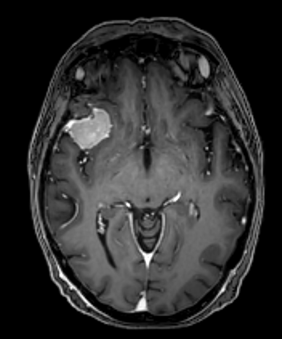
The gold standard for assessing sphenoid wing meningiomas is MRI imaging, which may be supplemented with CT imaging to assess the osseous structures, depending on the tumor. In selected cases, cerebral angiography may be indicated to facilitate preoperative embolization of the tumor’s vascular supply, if appropriate.

Treatment
Treatment of sphenoid wing meningiomas includes observation with regular follow-up imaging, microsurgical resection, Radiosurgery, and/or radiotherapy[^3].