Alongside the AICA and SCA, the PICA is one of the main vessels supplying the cerebellum.
Anatomy
The PICA usually arises directly from the Vertebral artery. It usually arises approximately 10 mm distal to the point at which the vertebral artery becomes intradural and approximately 15 mm proximal to the vertebrobasilar junction[^1]. In approximately 5-8 % of cases, the PICA has an extradural origin[^1]. Another anatomical variant is the AICA-PICA, in which the PICA arises from the Basilar artery at the same point as the AICA. On angiography, the PICA shows a cranial and a caudal loop, as well as the Choroidal Point. The PICA can be divided into five different segments[^4]:
Anterior medullary: From the origin of the PICA to the inferior aspect of the olive. Origin of 1 to 2 short medullary branches supplying the ventral medulla oblongata.
Lateral medullary: To the origin of cranial nerves IX, X & XI. Up to 5 perforators supplying the Brainstem.
Tonsillomedullary: To the tonsillar midportion. Includes the caudal loop on angiography.
Telovelotonsillary (supratonsillar): Ascends in the tonsillomedullary fissure. Includes the cranial loop on angiography.
Cortical segments
The first three segments must be preserved during a surgical procedure. The last two segments can be occluded with minimal deficits[^5].
Figure


Imaging


Clinical relevance
Aneurysms
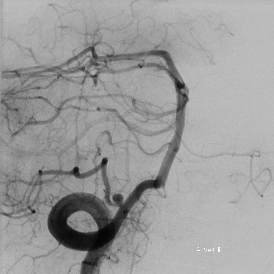
Aneurysms of the PICA account for 0,49 to 3 % of all cerebral aneurysms [^1] [^6].
Endoscopic endonasal approach for clipping of a PICA aneurysm
Operative video of an endoscopic endonasal approach for treatment of a PICA aneurysm.
Hemifacial spasm
In patients with Hemifacial spasm, the PICA is frequently the vessel responsible for the neurovascular conflict.
Neurological deficits
Occlusion of the PICA proximal to the Choroidal Point in the cranial loop carries a high risk of causing a medullary syndrome (e.g. Wallenberg syndrome), as a large number of medullary perforators arise proximal to this point[^3].