The anterior inferior cerebellar artery (AICA), along with the SCA and PICA, is one of the principal arterial vessels supplying the cerebellum.
Anatomy
Origin
The AICA usually originates directly from the Arteria basilaris. Normally, the AICA originates as a single trunk, but it may also consist of two or three trunks. The AICA may originate from any portion of the basilar artery, but usually from its caudal half [^1].
Course
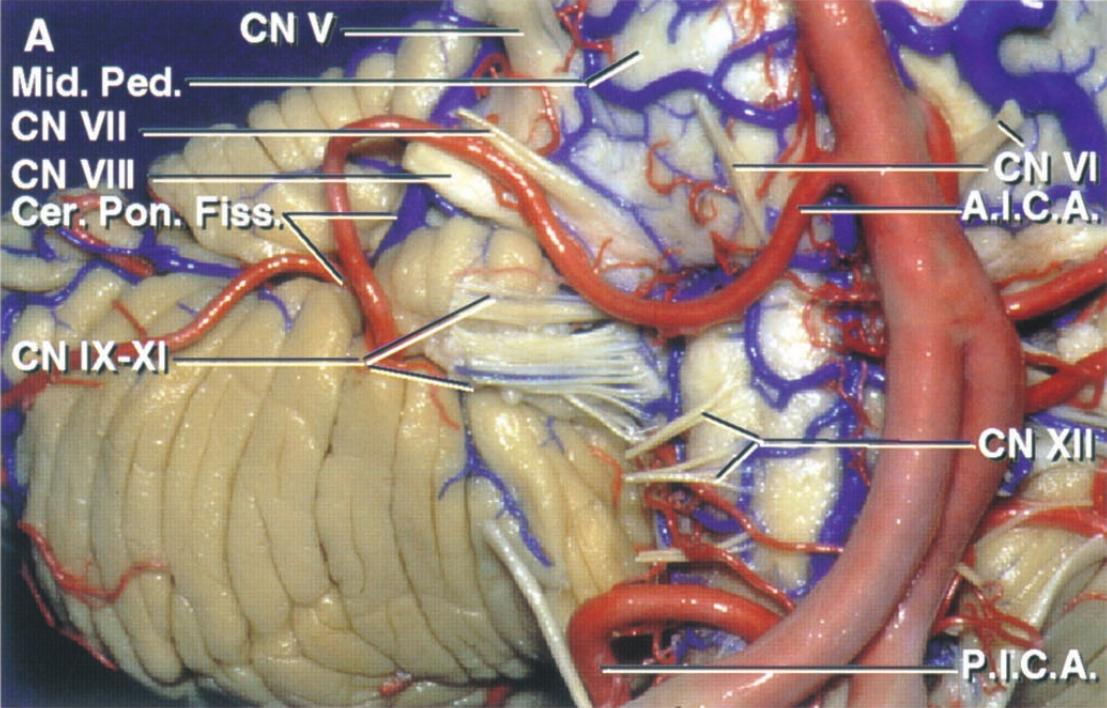
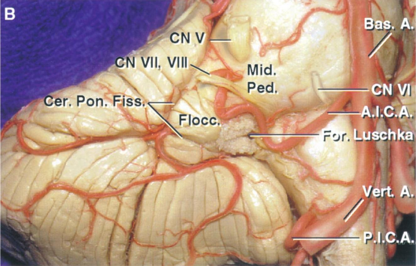
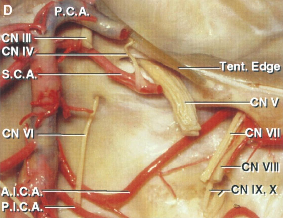
After its origin, the AICA courses around the pons in close proximity to the Abducens nerve, Facial nerve, and Vestibulocochlear nerve, and gives off smaller branches to the cranial nerves entering the internal acoustic meatus as well as to the Choroid plexus in the Foramen of Luschka. The AICA then courses around the flocculus and the middle cerebellar peduncle. Near the facial and vestibulocochlear nerves, the AICA frequently divides into a rostral and a caudal trunk. The rostral trunk gives off branches lateral to the middle cerebellar peduncle and to the cerebellopontine fissure and the surface of the petrous bone. The caudal trunk supplies the inferior portion of the surface of the petrous bone, the flocculus, and the choroid plexus. Along its course, the AICA gives off several perforating arteries to the brainstem as well as arteries accompanying nerves, such as the Labyrinthine artery [^1].
Segments
The AICA can be divided into four segments [^1]:
Anterior pontine segment
Lateral pontine segment
Flocculopeduncular segment
Cortical segment
Supply
The AICA supplies the anterolateral inferior portion of the Cerebellum and gives off the Arteria labyrinthi as another branch, which supplies the inner ear, the organ of hearing, and the organ of balance with arterial blood.
Figure