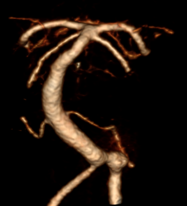
The vertebral artery is responsible for the main blood supply to the posterior circulation of the brain.
Anatomy
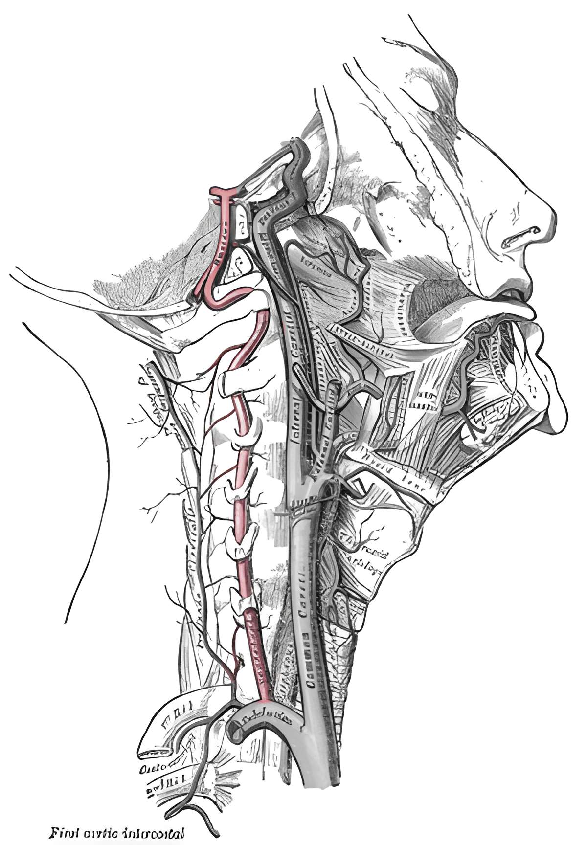
The paired vertebral arteries arise from the subclavian arteries and unite to form the Basilar artery. The vertebral artery can be divided into four segments[^1]:
Segments
Segment | Description | Arterial branches [^2] |
Prevertebral segment (V1) | The V1 segment extends from the arterial origin to its entry into the Foramen transversarium, usually at the level of C6 (HWK6) | None |
Foraminal segment (V2) | The V2 segment runs vertically through the foramina transversaria of the cervical spine. |
|
Predural segment (V3) | The V3 segment extends from the exit of the vertebral artery from the foramen transversarium of the axis to the entry of the vertebral artery into the Dura mater. |
|
Intradural segment (V4) | The V4 segment extends from the highly variable dural entry point to the point where the arteries unite to form the Basilar artery. |
Figure
Imaging
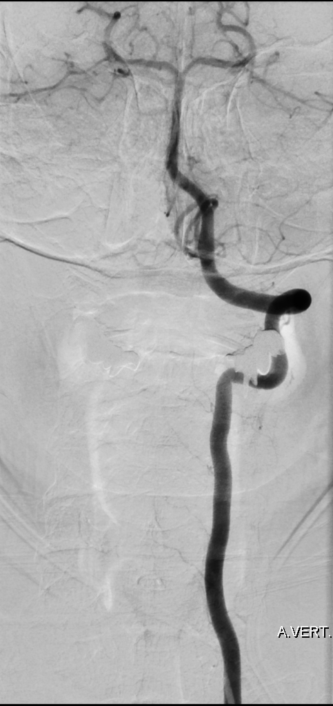
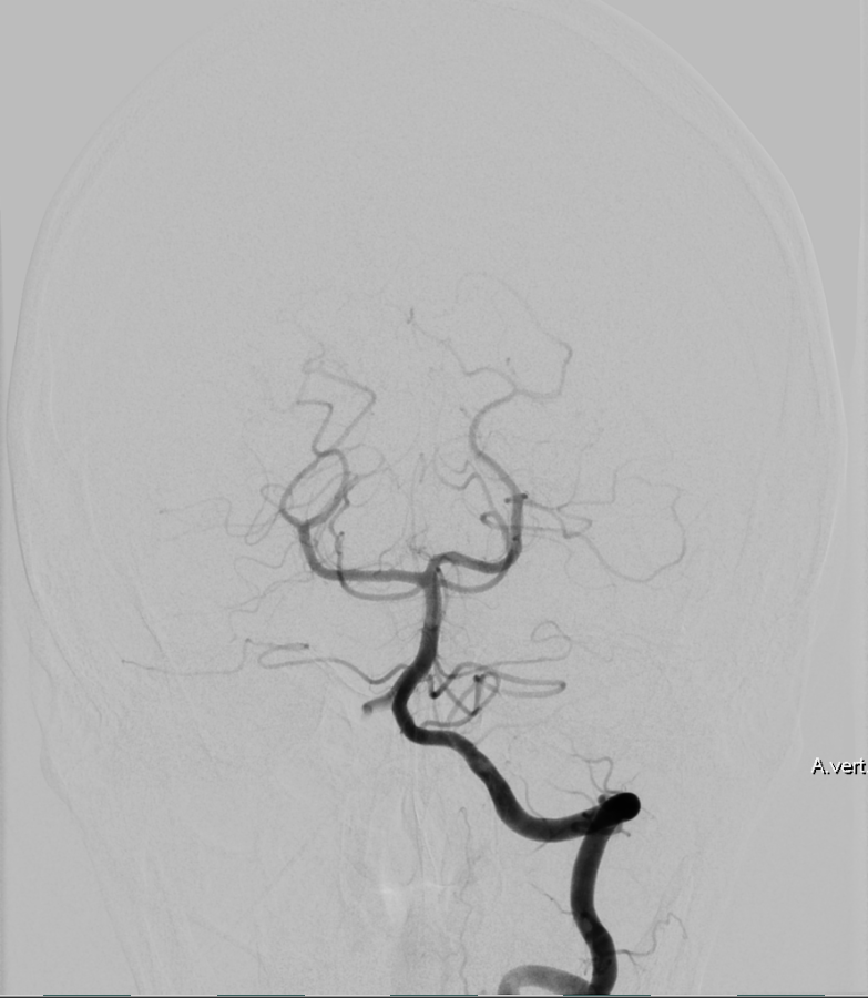
The gold standard for optimal visualization of the vertebral artery is digital subtraction angiography; however, depending on the clinical question, it can also be visualized using CT angiography or MRI imaging.

Clinical relevance
Aneurysms
Dissecting aneurysms can typically occur in the vertebral artery and cause a Subarachnoid hemorrhage[^3]. Depending on the case, these dissecting aneurysms may be treated endovascularly, for example by implantation of Flow diverters[^4] [^5].