Anterior Cervical Discectomy and Fusion (ACDF)

Synonyms
ACDF

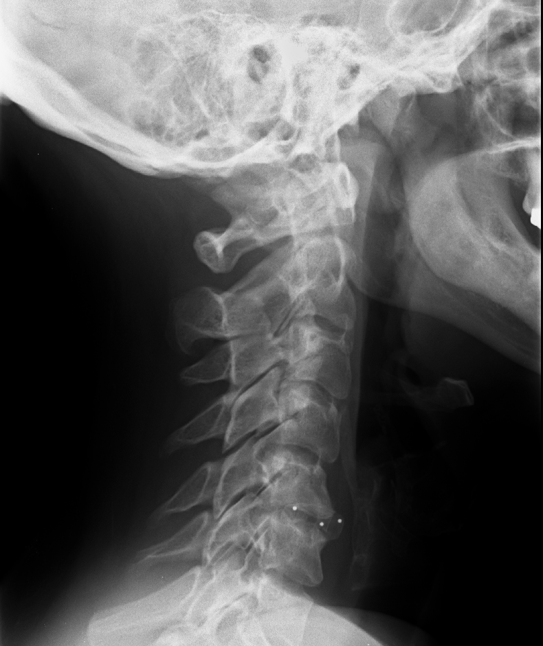
Anterior cervical discectomy and fusion (ACDF) is an established procedure for the treatment of cervical spine disorders, such as herniated discs, spinal canal stenosis, or advanced degenerative disc disease. This procedure enables removal of the intervertebral disc through an anterior approach, followed by fusion of the affected segment.

Plate

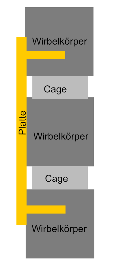
In some cases, additional placement of a plate is advisable or even necessary. When placing a plate, care should be taken to ensure that the plate does not cover more than 50 % of the last vertebral body affected by the plate in order to avoid adjacent osteophytes[^3]
Schematic ACDF plate
Schematic illustration of plating as part of an ACDF procedure.
ACDF plate
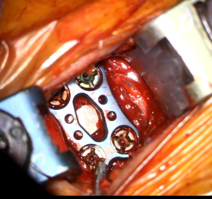
Intraoperative view of a single-level ACDF plate (Vectra) with a length of 16 mm.

Imaging

ACDF cage on radiograph
Visualization of a cage (red arrow) after an ACDF procedure.
Icotec cage ACDF radiograph
Postoperative radiograph after C5/6 ACDF with an Icotec cage.
Coalition Cage, cervical spine
Lateral radiograph of a patient with a Coalition Cage at the C5/6 level.

Complications

Complication rates for ACDF range from 13.2 % to 19.3 % in different studies[^2]. The most common complications include:

  • Dysphagia

  • postoperative hematomas

  • Aggravation of Myelopathy

  • symptomatic recurrent laryngeal nerve palsy

  • CSF leaks

  • wound infections

  • Aggravation of Radiculopathy

  • Horner syndrome

  • respiratory insufficiency

  • esophageal perforation

  • instrumentation failure

  • Pseudarthrosis occurs depending on the number of fused segments, ranging from 0 to 4.3 % for one segment to 56 % for four segments.

One of the most common long-term complications is adjacent segment degeneration, which involves degenerative changes in the segments adjacent to the surgical site. According to a study by Hilibrand et al. (1999), the risk of adjacent segment degeneration after ACDF surgery is approximately 2.9 % per year[^1].

Cage subsidence on X-ray
Lateral X-ray of a patient with a subsided cage
Cage Subsidence on CT
Sagittal CT image of a subsided cage after ACDF surgery.
Cage displacement on X-ray
Lateral X-ray image of a patient after an C5/6 ACDF procedure with a cage displaced anteriorly.

Editorial transparency

Authorship & revisions

Who created and updated this article, and which changes are documented.

Article created by
Last edited:
on
Revision history
8 edits · 1 contributor
Author list:
Show revision history 8 entries

8 edits by 1 author

Edit · Alaric Steinmetz · (+52 −26)

No content change recorded

Edit · Alaric Steinmetz ·

No content change recorded

Edit · Alaric Steinmetz · (+275 −0)

No content change recorded

Edit · Alaric Steinmetz · (+0 −269)

No content change recorded

Edit · Alaric Steinmetz ·

No content change recorded

Edit · Alaric Steinmetz · (+271 −0)

No content change recorded

Edit · Alaric Steinmetz · (+0 −275)

No content change recorded

Article created on · Alaric Steinmetz

Image viewer

Image

Open original