Posterior meningeal artery
Last edit by Alaric Steinmetz on
Synonyms: PMA, Posterior meningeal artery
The posterior meningeal artery (PMA) supplies parts of the dura mater.
Anatomy
The posterior meningeal artery originates from the ascending pharyngeal artery, a branch of the external carotid artery and enters intracranially via the jugular foramen [^1]. As an anatomical variant, the posterior meningeal artery can rarely also arise from the PICA [^2].
Imaging
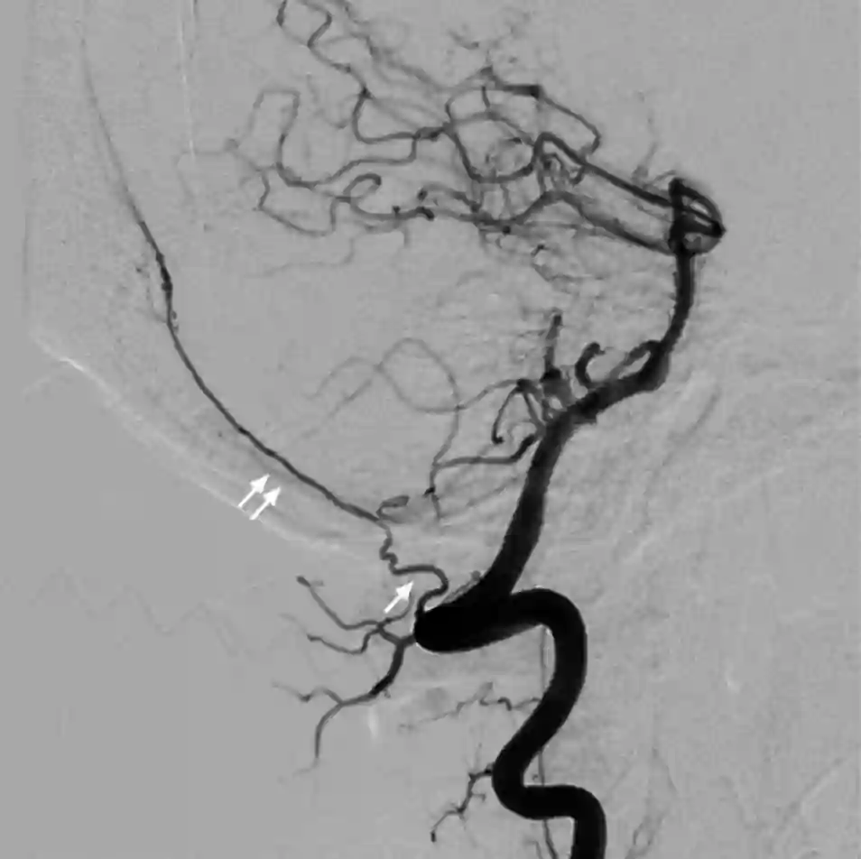
The posterior meningeal artery can be optimally demonstrated in digital subtraction angiography and can be divided into an extracranial and intracranial part. The extracranial part of the posterior meningeal artery is often coiled, whereas the intracranial part has a straight course [^3].

Clinical Relevance
The posterior meningeal artery is often involved in dural arteriovenous fistulas in the posterior fossa [^3].
In cases of ischemia or Moyamoya disease, the posterior meningeal artery can form anastomoses with pial arteries on the brain's surface, establishing a collateral circulation [^3].
Aneurysms can develop in the posterior meningeal artery, with the most common being traumatic pseudoaneurysms [^3].