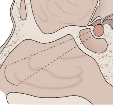
Tuberculum sellae meningiomas are Meningiomas that arise from the Tuberculum sellae.
Historical background
The first description of a Tuberculum sellae meningioma was provided by Stewart J. in 1899 in an autopsy report[^2].
Epidemiology
Tuberculum sellae meningiomas account for approximately 5-10 % of all intracranial meningiomas [^3].
Location
By definition, the anterior border of the Sulcus chiasmatis (Limbus sphenoidale) is the boundary between the anterior and middle cranial fossa. Therefore, Tuberculum sellae meningiomas are located in the middle cranial fossa. Planum sphenoidale- and olfactory meningiomas, in contrast, arise from the anterior cranial fossa.
Symptoms
The most common symptom of a symptomatic Tuberculum sellae meningioma is progressive, painless visual loss, which may be unilateral or bilateral, with homonymous or bitemporal hemianopia. Patients with larger Tuberculum sellae meningiomas may also develop endocrine dysfunction due to compression of the Pituitary gland or the Pituitary stalk. Less common symptoms include headache or epileptic seizures[^1].
Imaging
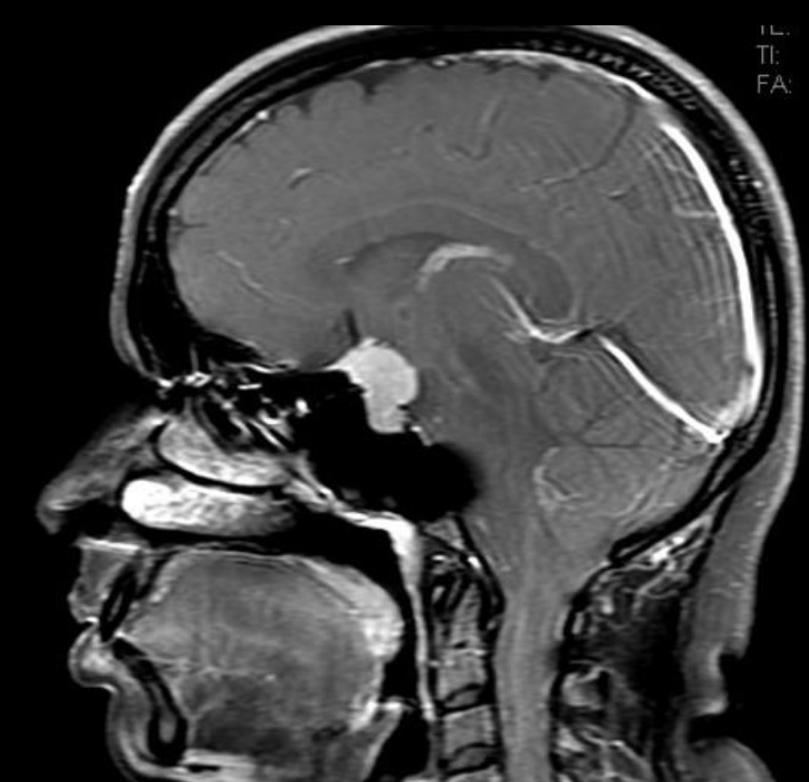
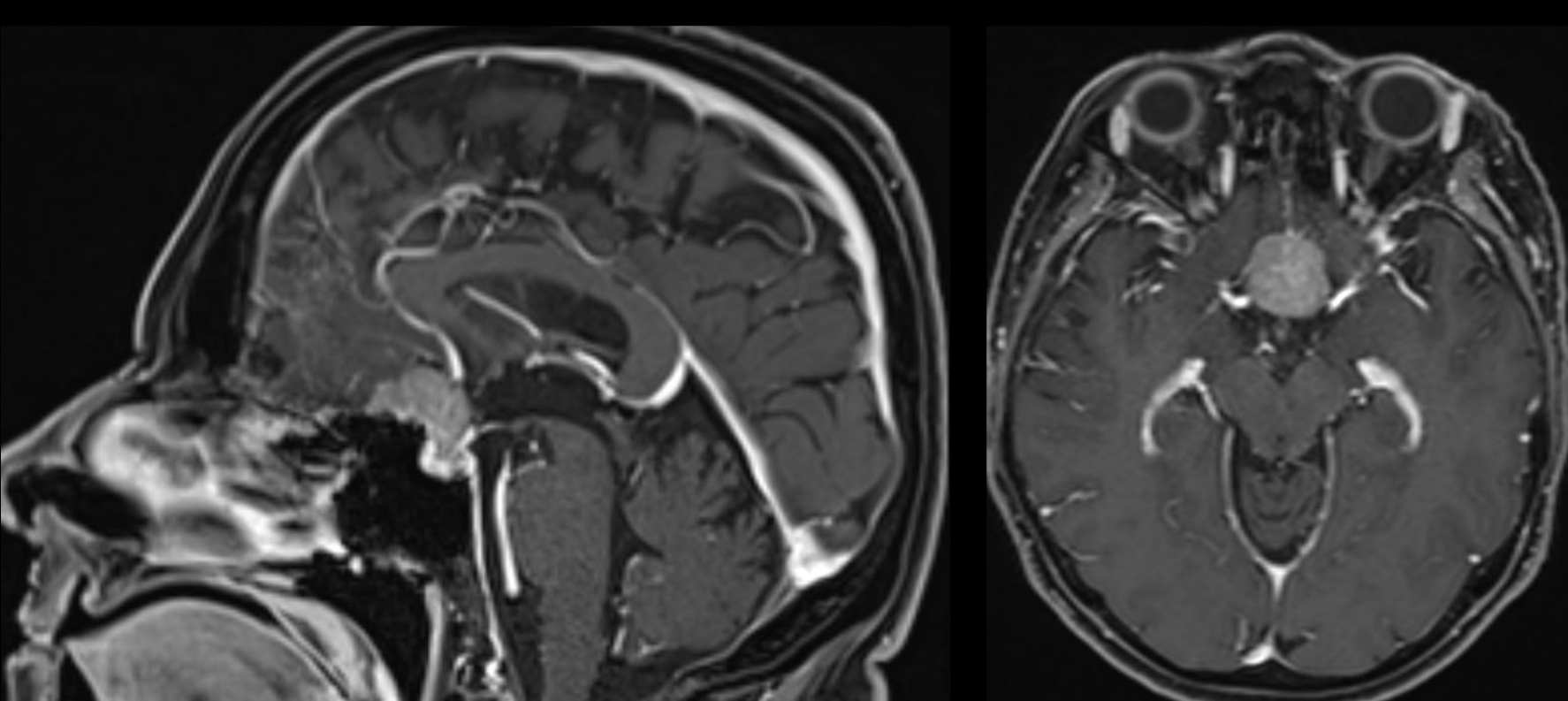
The gold standard for imaging Tuberculum sellae meningiomas is MRI imaging.
Surgical treatment
Tuberculum sellae meningiomas can be accessed and resected via different surgical approaches.
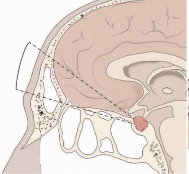
Unilateral subfrontal approach
A unilateral subfrontal approach is suitable for particularly large Tuberculum sellae meningiomas, as this approach provides optimal intraoperative visualization [^4].
Endoscopic endonasal approach
Another frequently used approach is the endoscopic endonasal approach [^4].