Transcranial Doppler Ultrasonography
Last edit by Alaric Steinmetz on
Synonyms: TCD
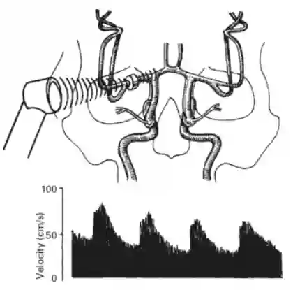
Transcranial Doppler ultrasonography is a procedure that can be performed at the patient's bedside to measure blood flow velocity in the cerebral arteries. By using low frequencies (~ 2 MHz), ultrasound can penetrate the temporal bone because the bone is generally very thin at this location1. The procedure is particularly suitable for measuring blood flow velocity in the large cerebral vessels, such as the anterior cerebral artery, middle cerebral artery, posterior cerebral artery, internal carotid artery, and basilar artery.
Application of transcranial Doppler ultrasonography in neurosurgery
In neurosurgery, the procedure is used particularly in patients with subarachnoid hemorrhage and is used here for the early diagnosis of vasospasm.

Acoustic windows
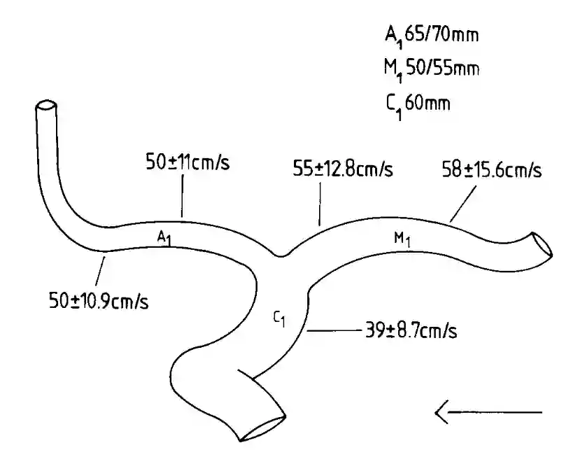
The M1, A1, and C1 segments can be assessed using transtemporal ultrasound.

Normal values
Normal values for the mean flow velocities (cm/s) in the middle, anterior, posterior, basilar, and vertebral vascular territories. Table adapted from Ringelstein et al.2:
| Age | MCA (M1) | ACA (A1) | PCA (P1) | BA | VA |
|---|---|---|---|---|---|
| 10-29 | 70 ± 16,4 | 61 ± 14,7 | 55 ± 9,0 | 46 ± 11 | 45 ± 9,8 |
| 30-49 | 57 ± 11,2 | 48 ± 7,1 | 42 ± 8,9 | 38 ± 8,6 | 35 ± 8,2 |
| 50-59 | 51 ± 9,7 | 46 ± 9,4 | 39 ± 9,9 | 32 ± 7,0 | 37 ± 10 |
| 60-70 | 41 ± 7,0 | 38 ± 5,6 | 36 ± 7,9 | 32 ± 6,7 | 35 ± 7,0 |
| Acoustic depth (mm) | 50-55 | 65-70 | 60-65 | 85-90 | 60-65 |
References
Lindsay, Kenneth W., Ian Bone, and Geraint Fuller. Neurology and neurosurgery illustrated e-book. Elsevier Health Sciences, 2010. ↩ ↩
Ringelstein, E. Bernd, et al. "Transcranial Doppler sonography: anatomical landmarks and normal velocity values." Ultrasound in medicine & biology 16.8 (1990): 745-761. ↩ ↩