Choroid plexus
Last edit by Alaric Steinmetz on
Synonyms: Plexus chorioideus, Choroid plexus, Plica choroidea
The choroid plexus is a knotted arteriovenous vascular convolutes in the brain ventricles, consisting of specialized glial cells and produce cerebrospinal fluid.
Anatomy
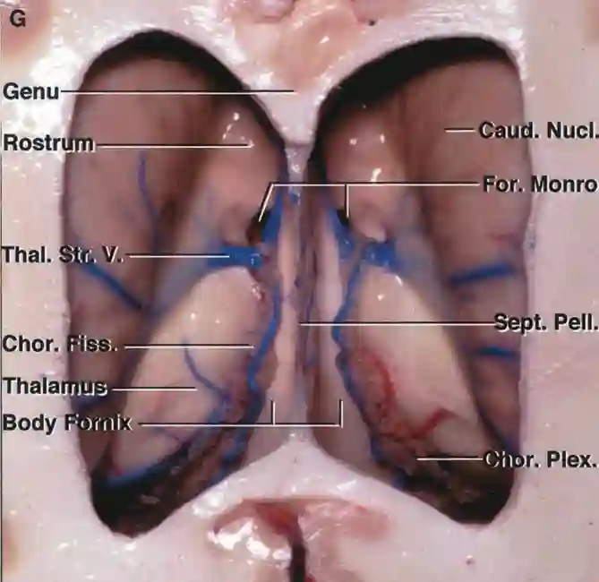
The choroid plexus is found in the lateral ventricles and in the roof of the third and fourth ventricles. At the lateral end of the fourth ventricle, a part of the choroid plexus protrudes bilaterally from the foramen of Luschka. This part is referred to as Bochdalek’s flower basket. The choroid plexus of the lateral ventricles is connected to that of the third ventricle via the interventricular foramen. Its attachment site at the thalamus is called the taenia choroidea.
Tumors
Tumors originating from the choroid plexus are called plexus tumors. In most cases, they are benign plexus papillomas (WHO Grade 1). Other more aggressive plexus tumors are the atypical plexus papilloma (WHO Grade 2) and the choroid plexus carcinoma (WHO Grade 3)[^1].
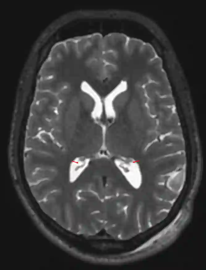
Imaging
The choroid plexus can be visible depending on its expression in CT and MRI imaging.

Figure