Nervus vestibulocochlearis

Synonyms
Vestibulocochlear nerve

The vestibulocochlear nerve is the eighth cranial nerve and consists of the vestibular nerve and the cochlear nerve.

Nuclei

The fibers of the vestibulocochlear nerve terminate in nuclei and are relayed from there:

  • Superior, medial, and inferior vestibular nuclei – to the vestibular organ

  • Lateral vestibular nuclei: receive most of the fiber components from the Purkinje cells of the cerebellum.

  • Anterior and posterior cochlear nuclei: to the auditory system

Imaging

The course of the vestibulocochlear nerve can, for example, be optimally visualized on a CISS sequence in MRI imaging.

Vestibulocochlear nerve on MRI
Depiction of the course of the vestibulocochlear nerve (marked in red) from the brainstem to the internal acoustic meatus on a T2 DRIVE MRI sequence.

Figure

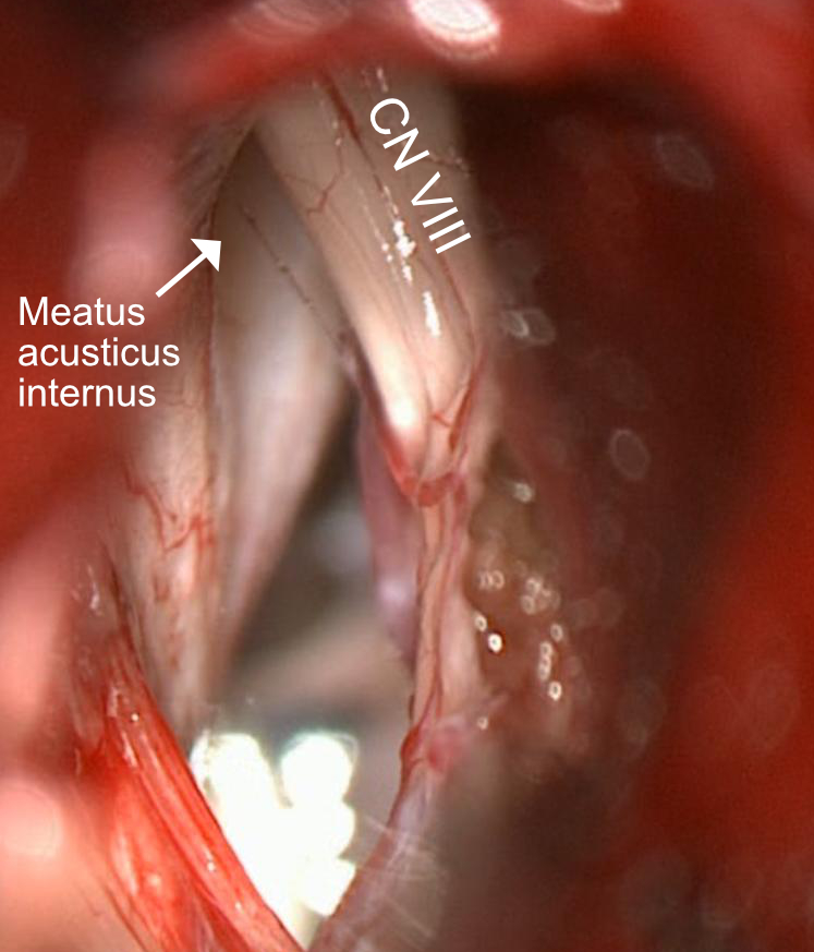
Retrosigmoid craniotomy
Depiction of the vestibulocochlear nerve (CN VIII) and the internal acoustic meatus during a retrosigmoid approach. The facial nerve is obscured by the vestibulocochlear nerve and is not visible.

Clinical relevance

A neurovascular conflict involving the vestibulocochlear nerve can lead to vestibular paroxysmia.

Editorial transparency

Authorship & revisions

Who created and updated this article, and which changes are documented.

Article created by
Last edited:
on
Revision history
4 edits · 1 contributor
Author list:
Show revision history 4 entries

4 edits by 1 author

Edit · Alaric Steinmetz ·

No content change recorded

Edit · Alaric Steinmetz · (+156 −0)

No content change recorded

Edit · Alaric Steinmetz · (+0 −154)

No content change recorded

Article created on · Alaric Steinmetz

Image viewer

Image

Open original