Acute subdural hematoma

Synonyms
Acute subdural hematoma, aSDH
ICD-10
S06.5

An acute subdural hematoma occurs in most cases after trauma1 and, depending on the extent of the hemorrhage, is considered a neurosurgical emergency requiring immediate treatment. In this condition, bleeding occurs between the brain and the dura mater.

Causes

In the vast majority of cases, an acute subdural hematoma results from direct traumatic impact to the skull and the resulting injury to the bridging veins2, leading to venous bleeding beneath the dura mater. As the bleeding increases, compression and displacement of the brain tissue toward the opposite side occur, resulting in a life-threatening situation.

In significantly rarer cases, an acute subdural hematoma can also develop atraumatically as a result of excessively rapid lumbar drainage of cerebrospinal fluid, such as with a lumbar drain3 or a surgically induced CSF leak[^3]. This can result in intracranial hypotension, which may cause injury to the bridging veins and thus lead to bleeding.

Diagnosis

The gold standard for diagnosing an acute subdural hematoma is CT imaging. However, the diagnosis can also be readily established with MRI.

Clinically, an acute subdural hematoma can manifest differently depending on its extent. When the hemorrhage has a significant mass effect, a decreased level of consciousness occurs in most cases, with considerable variation in severity. A hemisyndrome contralateral to the side of the hemorrhage is also typical. If the hematoma is very thin, it may also first become symptomatic in many cases through epileptic seizures.

Acute subdural hematoma on CT
An acute right-sided subdural hematoma on head CT.

Hyperacute subdural hematoma
Traumatic right-sided hyperacute subdural hematoma on CT.

Location

In theory, an acute subdural hematoma can occur anywhere in the subdural space. Traumatic subdural hematomas are most commonly found over the convexity, interhemispherically, and in the tentorial region.

Conservative treatment

In patients with very thin acute subdural hematomas without midline shift and without neurological deficits who are asymptomatic, conservative treatment without surgery may also be appropriate45. If conservative treatment is chosen, follow-up imaging is recommended to rule out further enlargement of the hematoma as well as chronicization.

Surgical treatment

Surgical evacuation of the hematoma is indicated in clinically symptomatic patients and in cases of extensive acute subdural hematomas with midline shift. This involves performing a craniotomy over the hematoma, opening the dura, and subsequently removing the hematoma. Normally, the removed bone flap is replaced. In situations in which markedly increased intracranial pressure is observed intraoperatively, the bone flap may also not be replaced, in the form of a decompressive hemicraniectomy6. The procedure is performed under general anesthesia.
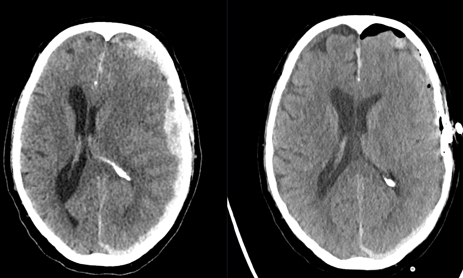
Acute subdural hematoma before and after surgery
Axial CT image of an acute left-sided subdural hematoma. Left: before surgery; right: after craniotomy and evacuation of the hematoma.

Prognostic outcome factors

Acute subdural hematoma is one of the most serious traumatic neurosurgical conditions. Patient outcomes depend on many factors. A significantly negative impact on outcome has been demonstrated for the following factors: motorcycle accident as the mechanism of injury, patient age over 65, a GCS of 3 to 4 on arrival at the trauma center, and a postoperative ICP of > 45 mmHg7.

In patients with a GCS from 3 to 7, mortality was 66 % and functional recovery was 19 % among those who required surgery because of an acute subdural hematoma8.

In the study by Wilberger et al. from 19919, outcomes were stratified according to initial GCS and age in 101 patients as follows.

Outcome stratification by GCS

GCS Outcome mortality10 Functional recovery11
3 90 % 5 %
4 76 % 10 %
5 62 % 18 %
6-7 51 % 44 %

Outcome stratification by age

Stratification of outcomes by age in patients with an initial GCS of 3 to 712.

Age Outcome mortality Functional recovery
34 54,3 % 31,4 %
35-50 61,9 % 23,8 %
51-65 70,6 % 11,8 %
>65 82,1 % 3,6 %

Time to surgery also plays an important prognostic role. In 1981, it was shown that patients with an acute subdural hematoma who underwent surgery within 4 hours after trauma had a mortality of 30 %, whereas patients who required more than 4 hours to reach surgery had a mortality of 90 %13.

References


    Editorial transparency

    Authorship & revisions

    Who created and updated this article, and which changes are documented.

    Article created by
    Last edited:
    on
    Revision history
    4 edits · 1 contributor
    Author list:
    Show revision history 4 entries

    4 edits by 1 author

    Edit · Alaric Steinmetz · (+4 −4)

    No content change recorded

    Edit · Alaric Steinmetz ·

    No content change recorded

    Edit · Alaric Steinmetz · (+24 −24)
    • Text changed: “In most cases, an acute subdural hematoma occurs after trauma[^1] and, depending on the extent of the blood volume, is considered a neurosurgical emergency requiring immediate treatment. This involves bleeding between the brain and the dura mater.”“An acute subdural hematoma occurs in most cases after trauma[^1] and, depending on the extent of the hemorrhage, is considered a neurosurgical emergency requiring immediate treatment. In this condition, bleeding occurs between the brain and the dura mater.”
    • Text changed: “In the vast majority of cases, an acute subdural hematoma develops as a result of direct traumatic impact on the skull and consequent damage to the bridging veins[^1], followed by venous bleeding beneath the [dura mater](\/lexikon\/dura-mater). As the bleeding increases, compression and displacement of the brain tissue toward the opposite side occur, resulting in a life-threatening situation.”“In the vast majority of cases, an acute subdural hematoma results from direct traumatic impact to the skull and the resulting injury to the bridging veins[^1], leading to venous bleeding beneath the [dura mater](\/lexikon\/dura-mater). As the bleeding increases, compression and displacement of the brain tissue toward the opposite side occur, resulting in a life-threatening situation.”
    • Text changed: “In significantly rarer cases, an acute subdural hematoma can also develop atraumatically as a result of overly rapid lumbar drainage of cerebrospinal fluid, such as with a [lumbar drain](\/lexikon\/lumbaldrainage)[^2] or a surgically induced [CSF leak](\/lexikon\/liquorfistel)[^3]. This results in intracranial hypotension, which can cause damage to the bridging veins and consequently lead to bleeding.”“In significantly rarer cases, an acute subdural hematoma can also develop atraumatically as a result of excessively rapid lumbar drainage of cerebrospinal fluid, such as with a [lumbar drain](\/lexikon\/lumbaldrainage)[^2] or a surgically induced [CSF leak](\/lexikon\/liquorfistel)[^3]. This can result in intracranial hypotension, which may cause injury to the bridging veins and thus lead to bleeding.”
    • Text changed: “The gold standard in the diagnosis of an acute subdural hematoma is CT imaging. However, the diagnosis can also be readily made on MRI.”“The gold standard for diagnosing an acute subdural hematoma is CT imaging. However, the diagnosis can also be readily established with MRI.”
    • Text changed: “Clinically, an acute subdural hematoma can manifest differently depending on its extent. In most cases, a markedly space-occupying hemorrhage results in a reduction in level of consciousness, which may vary considerably in severity. A hemiparesis contralateral to the side of the hemorrhage is also typical. If the hematoma is very thin, it may in many cases first become symptomatic through epileptic seizures.”“Clinically, an acute subdural hematoma can manifest differently depending on its extent. When the hemorrhage has a significant mass effect, a decreased level of consciousness occurs in most cases, with considerable variation in severity. A hemisyndrome contralateral to the side of the hemorrhage is also typical. If the hematoma is very thin, it may also first become symptomatic in many cases through epileptic seizures.”
    • Text changed: “An acute subdural hematoma can theoretically occur anywhere in the subdural space. Traumatic subdural hematomas are most commonly found over the convexity, interhemispherically, and tentorially.”“In theory, an acute subdural hematoma can occur anywhere in the subdural space. Traumatic subdural hematomas are most commonly found over the convexity, interhemispherically, and in the tentorial region.”
    • Text changed: “## Conservative Treatment {#konservative-therapie}”“## Conservative treatment {#konservative-therapie}”
    • Text changed: “In cases of very thin acute subdural hematomas without midline shift, as well as in patients without neurological deficits who are asymptomatic, conservative treatment without surgery may also be pursued[^4][^5]. If conservative treatment is chosen, follow-up imaging is recommended to rule out further enlargement of the hematoma as well as chronic transformation.”“In patients with very thin acute subdural hematomas without midline shift and without neurological deficits who are asymptomatic, conservative treatment without surgery may also be appropriate[^4][^5]. If conservative treatment is chosen, follow-up imaging is recommended to rule out further enlargement of the hematoma as well as chronicization.”
    • Text changed: “## Surgical Management {#operative-versorgung}”“## Surgical treatment {#operative-versorgung}”
    • Text changed: “Surgical removal of the hematoma is indicated in clinically symptomatic patients and in cases of extensive acute subdural hematomas with midline shift. This involves a craniotomy over the hematoma and opening of the dura, followed by removal of the hematoma. Normally, the removed bone flap is replaced. In situations in which markedly elevated intracranial pressure is observed intraoperatively, the bone flap may also not be replaced, constituting a [decompressive hemicraniectomy](\/lexikon\/kraniektomie)[^6]. The procedure is performed under general anesthesia.”“Surgical evacuation of the hematoma is indicated in clinically symptomatic patients and in cases of extensive acute subdural hematomas with midline shift. This involves performing a craniotomy over the hematoma, opening the dura, and subsequently removing the hematoma. Normally, the removed bone flap is replaced. In situations in which markedly increased intracranial pressure is observed intraoperatively, the bone flap may also not be replaced, in the form of a [decompressive hemicraniectomy](\/lexikon\/kraniektomie)[^6]. The procedure is performed under general anesthesia.”
    • Text changed: “## Prognostic Outcome Factors {#prognose-faktoren}”“## Prognostic outcome factors {#prognose-faktoren}”
    • Text changed: “Acute subdural hematoma is one of the most severe traumatic neurosurgical conditions. Patient outcome depends on many factors. A significantly negative impact on outcome has been demonstrated for the following factors: motorcycle accident as the mechanism of injury, patient age over 65, [GCS](\/scores-und-klassifikationen\/glasgow-coma-scale) of 3 to 4 on arrival at the trauma center, and postoperative [ICP](\/lexikon\/intrakranieller-druck-icp) of > 45 mmHg[^7].”“Acute subdural hematoma is one of the most serious traumatic neurosurgical conditions. Patient outcomes depend on many factors. A significantly negative impact on outcome has been demonstrated for the following factors: motorcycle accident as the mechanism of injury, patient age over 65, a [GCS](\/scores-und-klassifikationen\/glasgow-coma-scale) of 3 to 4 on arrival at the trauma center, and a postoperative [ICP](\/lexikon\/intrakranieller-druck-icp) of > 45 mmHg[^7].”
    • Text changed: “From 3 to 73-7, a mortality of 66 % and functional recovery of 19 % were observed in patients who had to undergo surgery because of an acute subdural hematoma[^7].”“In patients with a GCS from 3 to 7, mortality was 66 % and functional recovery was 19 % among those who required surgery because of an acute subdural hematoma[^7].”
    • Text changed: “In the study by Wilberger et al. from 1991[^7], outcome was stratified according to initial GCS and age in 101 patients as follows.”“In the study by Wilberger et al. from 1991[^7], outcomes were stratified according to initial GCS and age in 101 patients as follows.”
    • Text changed: “### Outcome Stratification by GCS {#outcome-stratifizierung-gcs}”“### Outcome stratification by GCS {#outcome-stratifizierung-gcs}”
    • Text changed: “| GCS | Outcome Mortality[^7] | Functional Recovery[^7] | | -------- | ----------|------------- | | 3 | 90 % | 5 % | | 4 | 76 % | 10 % | | 5 | 62 % | 18 % | | 6-7 | 51 % | 44 % |”“| GCS | Outcome mortality[^7] | Functional recovery[^7] | | -------- | ----------|------------- | | 3 | 90 % | 5 % | | 4 | 76 % | 10 % | | 5 | 62 % | 18 % | | 6-7 | 51 % | 44 % |”
    • Text changed: “### Outcome Stratification by Age {#outcome-stratifizierung-alter}”“### Outcome stratification by age {#outcome-stratifizierung-alter}”
    • Text changed: “Stratification of outcome by age for an initial GCS of 3 to 7[^7].”“Stratification of outcomes by age in patients with an initial GCS of 3 to 7[^7].”
    • Text changed: “| Age | Outcome Mortality | Functional Recovery | | -------- | ----------|------------- | | 34 | 54,3 % | 31,4 % | | 35-50 | 61,9 % | 23,8 % | | 51-65 | 70,6 % | 11,8 % | | >65 | 82,1 % | 3,6 % |”“| Age | Outcome mortality | Functional recovery | | -------- | ----------|------------- | | 34 | 54,3 % | 31,4 % | | 35-50 | 61,9 % | 23,8 % | | 51-65 | 70,6 % | 11,8 % | | >65 | 82,1 % | 3,6 % |”
    • Text changed: “Time to surgery also plays an important prognostic role. In 1981, it was shown that patients who underwent surgery for an acute subdural hematoma within 4 hours of the trauma had a mortality of 30 %, whereas patients who required more than 4 hours before surgery had a mortality of 90 %[^8].”“Time to surgery also plays an important prognostic role. In 1981, it was shown that patients with an acute subdural hematoma who underwent surgery within 4 hours after trauma had a mortality of 30 %, whereas patients who required more than 4 hours to reach surgery had a mortality of 90 %[^8].”
    • Image added: interhempherisches_subduralhaematom.png

    Article created on · Alaric Steinmetz

    Image viewer

    Image

    Open original