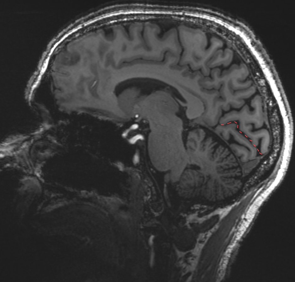
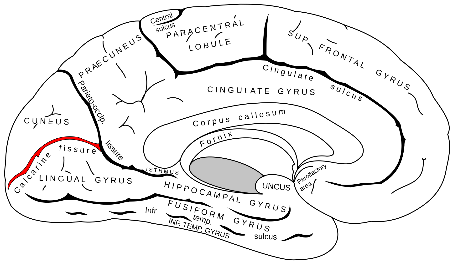
The sulcus calcarinus is a Sulcus of the medial occipital lobe.
Anatomy
The sulcus calcarinus begins above the splenium of the Corpus callosum and caudally separates the isthmus of the Cingulate gyrus from the Parahippocampal gyrus. Posteriorly, the sulcus calcarinus follows a gentle, upwardly convex curve, from the apex of which the Parieto-occipital sulcus arises superiorly; this, in turn, delineates the occipital lobe anteriorly on the medial surface of the cerebral hemisphere. Posteriorly, the sulcus calcarinus may occasionally cross the superomedial margin and extend along the Occipital pole to the superolateral surface of the cerebral hemisphere.
The point at which the parieto-occipital sulcus arises divides the sulcus calcarinus into a proximal and a distal part. Superior to the proximal half of the sulcus calcarinus and anterior to the parieto-occipital sulcus lies the Precuneus, which is part of the parietal lobe. Superior to the distal part and posterior to the parieto-occipital sulcus is the Cuneus[^1]. The calcarine branch of the medial occipital artery runs within the sulcus calcarinus.
Figure
Imaging
Macroscopically and on imaging, the sulcus calcarinus is best visualized in the midsagittal section of the brain.