Ependymomas are usually slow-growing tumors of the central nervous system that morphologically resemble the Ventricular system and the Spinal canal-lining Ependymal cells.
Epidemiology
According to the "Central Brain Tumor Registry of the United States," the annual incidence of ependymal tumors is approximately 0.43 patients per 100,000 inhabitants. Ependymomas account for approximately 1.8 % of all primary CNS tumors and approximately 6.8 % of all gliomas. In children and adolescents (0-19 years), ependymal tumors occur proportionally more frequently and account for approximately 5.2 % of all primary CNS tumors. Males are affected more frequently than females (1.3:1)[^2]. Although ependymomas occur preferentially in childhood and adolescence, they can occur at any age[^1].
Classification
According to the 2021 WHO CNS classification, supratentorial and infratentorial ependymomas are classified as WHO Grade 2 or 3[^5].
Location
Two-thirds of ependymomas occur infratentorially in the Fourth ventricle; the remaining third is located supratentorially, usually in the periventricular white matter. In contrast to a Medulloblastoma, which frequently arises from the roof of the fourth ventricle, an ependymoma often spreads from the floor of the fourth ventricle.
Imaging
On imaging, ependymomas differ from medulloblastomas primarily in their frequent calcification, which occurs in fewer than 10 % of medulloblastoma cases[^2]. Ependymomas also frequently appear heterogeneous on T1-weighted sequences, and the exophytic component of ependymomas is often hyperintense on T2-weighted sequences[^2]. Ependymomas may show restricted diffusion on MRI, although this occurs significantly less frequently in ependymomas than in medulloblastomas[^4].
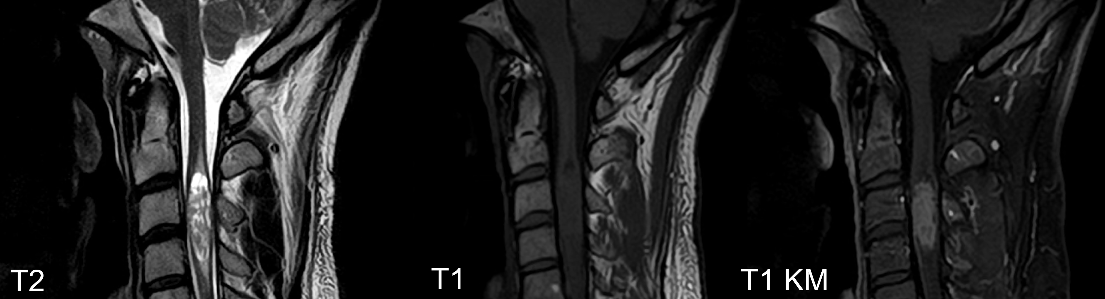
A possible, but not definitive, distinguishing feature between spinal ependymomas and astrocytomas is the presence of Syringohydromyelia in ependymomas, which occurs only very rarely in astrocytomas[^7].


Intramedullary ependymomas
Ependymomas can occur along the entire spinal cord and are the most common gliomas of the caudal spinal cord, Conus, and Filum. They are slow-growing tumors.

Surgical videos
Resection of intradural intramedullary subependymoma of the cervicothoracic spine: 2-dimensional operative video
Operative video of the resection of an intramedullary ependymoma.
Spinal metastases
The incidence of spinal metastases at the initial diagnosis of an ependymoma is approximately 5 %[^3].
Treatment guidelines
The current guidelines and treatment recommendations for ependymomas can be found here: EANO Guidelines.