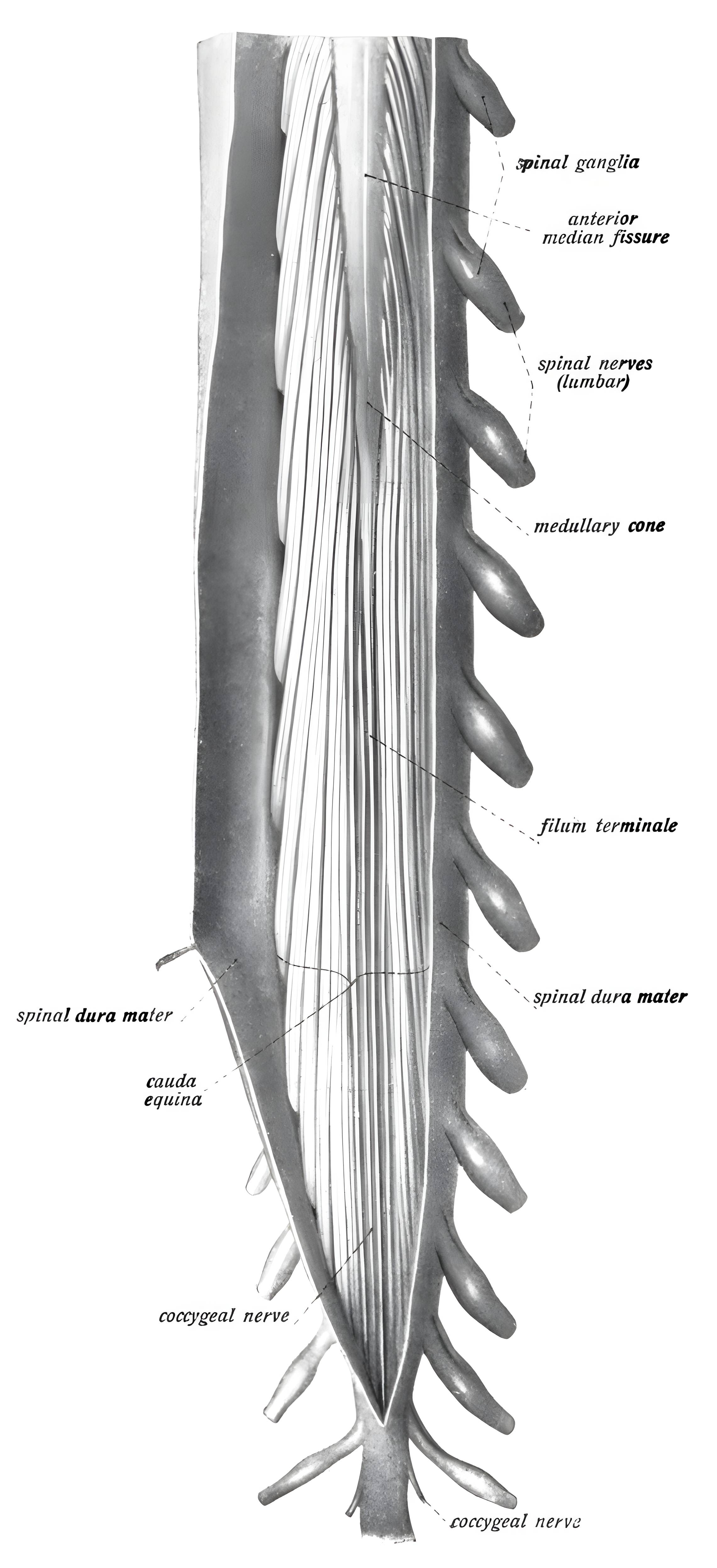
The filum terminale, formerly referred to as the Nervus impar, is a thin strand of connective tissue that extends caudally from the conus medullaris of the spinal cord. The filum terminale is divided into an internal filum terminale and an external filum terminale.
Internal filum terminale
This is an approximately 15 cm-long strand of connective tissue. It extends approximately to the level of the second sacral vertebra. As an extension of the pia mater spinalis, it lies within the dural sac and is surrounded by the nerve roots of the cauda equina.
External filum terminale
The lower segment is referred to as the external filum terminale. Here, the tissue fuses with the dura mater spinalis and forms a tough strand that ultimately attaches to the first coccygeal vertebra. The external filum terminale also contains smooth muscle cells1.
Intraoperative differentiation
The intraoperative differentiation of the filum terminale from a nerve root can be performed as follows:
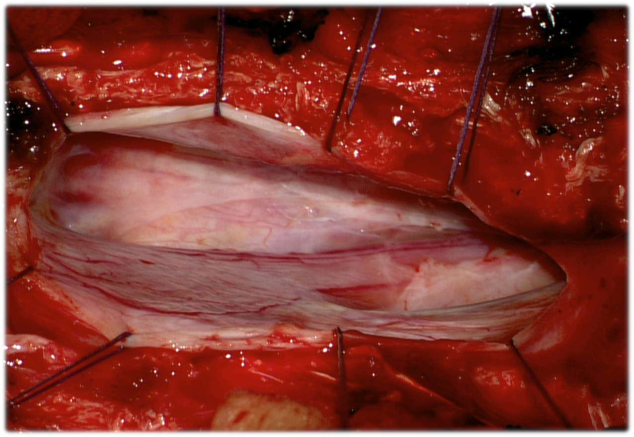
- A tortuous vessel often runs along the surface of the filum terminale2.
- Under the microscope, the filum terminale appears whiter than nerve roots, and ligamentous fibers may be partially observed3.
- Intraoperative electrical stimulation and EMG recordings of the anal sphincter musculature provide greater certainty in distinguishing between them4.
Figure
Function
The filum terminale prevents rostral migration of the spinal cord5.
References
Tubbs, R. S., Murphy, R. L., Kelly, D. R., Lott, R., Salter, E. G., & Oakes, W. J. (2005). The filum terminale externum, Journal of Neurosurgery: Spine, 3(2), 149-152. ↩ ↩
Handbook of Neurosurgery. Greenberg M, ed. 9th Edition. New York: Thieme ↩ ↩ ↩ ↩ ↩ ↩
Book: Neurosurgical Knowledge Update - A comprehensive review. Robert E. Harbaugh et al. Thieme Verlag. ↩ ↩