Subependymomas are slow-growing tumors and are classified as WHO Grade 1 tumors.
Epidemiology
Subependymomas account for 0.51 % of all tumors of the central nervous system and approximately 8 % of all ependymal tumors[^1] [^2].
Etiology
Subependymomas arise from the Ependymal cells in the Ventricles, protrude into the ventricular system, and grow noninvasively.
Diagnosis
Subependymomas are usually incidental imaging findings or become symptomatic due to symptoms of Hydrocephalus. Subependymomas can occur in the Brain as well as the Spinal cord.
Imaging
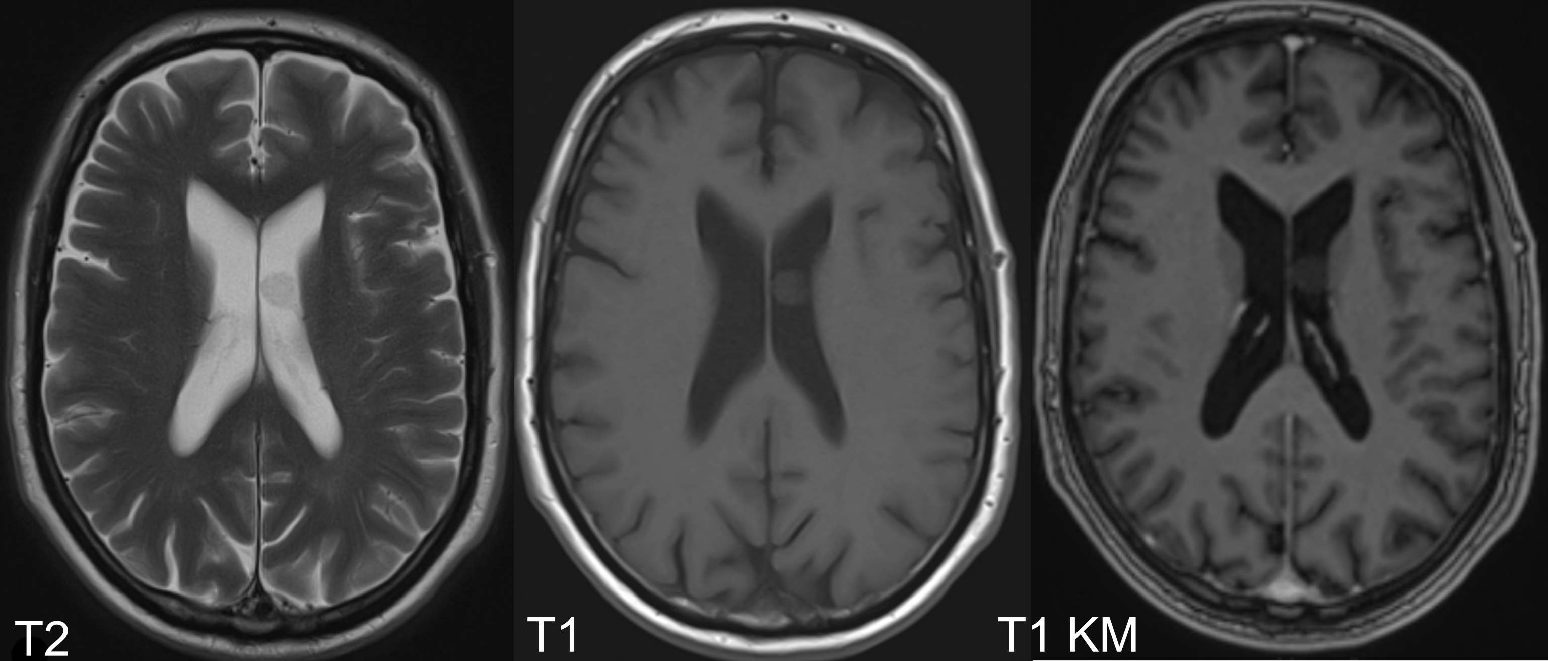
The gold standard for imaging subependymomas is MRI imaging. On MRI, subependymomas appear as hypo- to hyperintense lesions on T1- and T2-weighted sequences and typically show no or only minimal contrast enhancement[^1] [^2].

Treatment
Subependymomas are slow-growing tumors and can be monitored with follow-up imaging if asymptomatic. If surgical removal is indicated, gross total resection (GTR) is usually curative, and subtotal resection is generally also sufficient due to the slow tumor growth[^1].