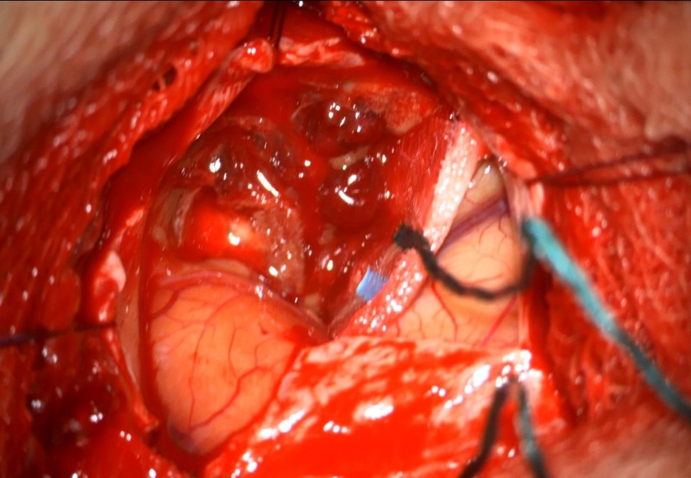
Cavernous malformations, also known as cavernomas, are vascular malformations with a sinusoidal architecture that are lined only by endothelium with irregularly thick walls. Cavernous malformations do not have large feeding arteries or large draining veins. In most cases, cavernous malformations have a diameter of 1-5 cm and may calcify, thrombose, or hemorrhage[^1].

Epidemiology
The average age at which cavernous malformations are diagnosed is approximately 30 years, and they are diagnosed with equal frequency in women and men[^2]. Cavernous malformations account for approximately 5-13 % of all vascular pathologies in the central nervous system and develop in approximately 0,02-0,16 % of the population[^1]. 48-86 % of cavernous malformations are located supratentorial, 4-35 % in the brainstem, and approximately 5-10% in the basal ganglia[^3]. Multiple cavernous malformations are present at diagnosis in 23 %[^4] to 50 %[^5] of cases. In very rare cases, cavernous malformations can also occur in the spinal cord. Cavernous malformations may also occur with familial clustering, for which several genetic loci have been identified: CCM1, CCM2, and CCM3. The PIK3CA locus has been identified for sporadic cavernous malformations[^9].
Symptoms
Cavernous malformations become symptomatic in 50 % of cases through epileptic seizures or symptoms of acute hemorrhage from the cavernous malformation. Patients frequently present with acute-onset headaches or new neurological deficits that correspond to the location of the cavernous malformation. A first hemorrhage from a cavernous malformation is rarely fatal. The risk of epileptic seizures is particularly high with supratentorial cavernous malformations.
Imaging
The gold standard for diagnosing cavernous malformations is MRI imaging.


Cavernomatosis on MRI
SWI MRI sequence of a patient with extensive cavernomatosis.
Risk of hemorrhage
The risk of acute hemorrhage depends on the location of the cavernous malformation. Cavernous malformations located deep or in the brainstem have an annual hemorrhage risk of approximately 4-5 % per lesion each year. Superficially located supratentorial cavernous malformations have an annual hemorrhage risk of <1 %[^6].
Location
Spinal intramedullary cavernous malformations
Intramedullary cavernous malformations may behave clinically and radiologically very similarly to demyelinating or neoplastic processes[^8]. Approximately 27 % of patients also have concurrent cranial cavernous malformations. Spinal cavernous malformations may present with varying clinical manifestations. Approximately 30 % of patients experience stepwise deterioration, approximately 41 % progressive deterioration, and approximately 26 % acute neurological deterioration[^7].
Diagnosis
Cavernous malformations are usually diagnosed with MRI imaging, with T2 and SWI sequence being particularly helpful. On MRI, cavernous malformations typically show a "popcorn-like" or "mulberry-like" appearance. They are frequently classified according to the Zabramski classification. digital subtraction angiography does not visualize cavernous malformations, but may play a role in the diagnostic workup to exclude other vascular pathologies in unclear cases[^1].
Prognosis
Following complete surgical removal, the risk of regrowth or hemorrhage is generally completely eliminated[^1]. However, symptoms may recur after partial removal or even after presumed complete removal[^1]. Postoperatively, patients with cavernous malformations, particularly those located in the brainstem, may frequently experience postoperative deterioration, which is generally transient but may persist for months[^1].