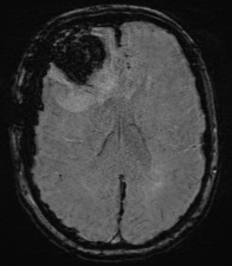
Susceptibility-weighted imaging (Susceptibility-Weighted Imaging, SWI) is an MRI imaging technique used in medical diagnostics, specifically for the venous vasculature.
Historical background
The underlying principle was first published in 1997[^1] and described comprehensively in 2001[^2].
Background
The SWI sequence is based on the physical property of magnetic susceptibility. SWI is a magnetic resonance imaging technique. It uses flow-compensated, spatially high-resolution 3D gradient-echo sequences (GRE sequence) in single-echo and multiecho techniques, taking advantage of the different magnetic susceptibilities of the various tissues. These differences lead to a phase difference and result in signal loss. No contrast agent is used. Combining the magnitude and phase images produces an enhanced contrast image that can depict venous blood, (cerebral) hemorrhages, and iron deposits such as hemosiderin. Imaging of venous blood with SWI is referred to as blood oxygen level-dependent imaging (BOLD, blood-oxygen-level dependent). Venous (deoxygenated) blood is less diamagnetic than arterial (oxygenated) blood. The technique was therefore initially referred to as BOLD, but was later replaced by the more general term susceptibility-weighted imaging. The term BOLD venography is still occasionally used today. Due to the BOLD effect, SWI provides good visualization of the venous vasculature.
Applications
SWI sequences may be used, for example, in patients with Traumatic brain injury, for high-resolution cerebral venography, and in other clinical applications.