Zabramski Classification
Last edit by Alaric Steinmetz on
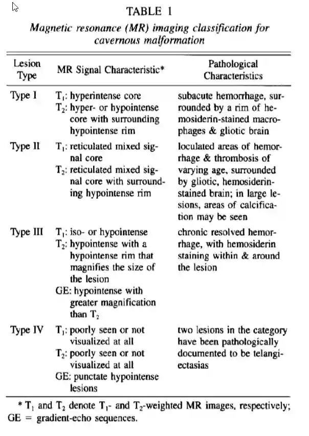
The Zabramski classification was published in 1994 by Zabramski et al.[^1] and is used for the neuroradiological classification of cavernomas.
Classification
Zabramski type | MRI gradient echo signal | Pathological characteristics | ||
Type I | Hyperintense core | Hyper- or hypointense core with a hypointense rim | - | Subacute hemorrhage surrounded by a rim of hemosiderin, macrophages, and gliotic brain tissue. |
Type II | Reticular mixed core | Reticular mixed core with a hypointense rim | - | Areas of hemorrhage and thrombosis of varying ages surrounded by hemosiderin and gliotic brain tissue. Calcifications may occur in part. |
Type III | Iso- or hypointense | Hypointense with a hypointense rim that enlarges the lesion. | Hypointense, but larger than in the T2 sequence. | Chronified blood with hemosiderin in the hemorrhage and at the margin. |
Type IV | Not or barely visible | Not or barely visible | Punctate hypointense lesions. | Two lesions in this category were documented as telangiectasias in the original study. |
Figure