The pineal cyst is a benign cystic mass in the area of the Pineal gland. Over 80% of pineal cysts are less than 10 mm in size and very rarely show progression in follow-up imaging.
Epidemiology
Pineal cysts are found in 1.8% to 4.3% of all cranial MRI scans and therefore very often occur as asymptomatic incidental findings[^1][^8]. Autopsy studies suggest that a pineal cyst is present in up to 40% of the population[^9] [^10].
Symptoms
Larger pineal cysts can lead to compression of the aqueduct and a consequent hydrocephalus.
Imaging
Depending on their manifestation and size, pineal cysts can be visible in both CT and MRI imaging. The gold standard for imaging depiction is MRI.
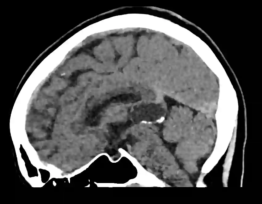
CT
Small pineal cysts can often be overlooked on CT imaging due to their similar intensity to cerebrospinal fluid.

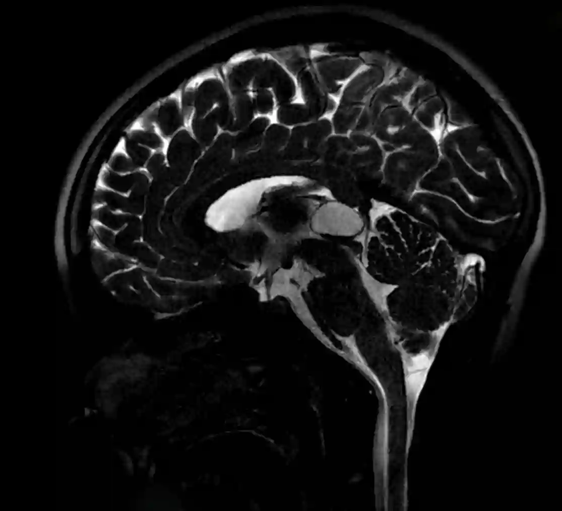
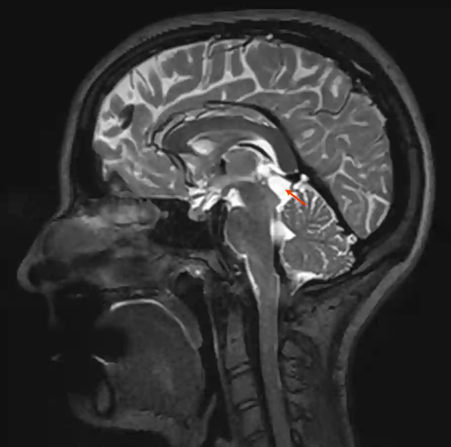
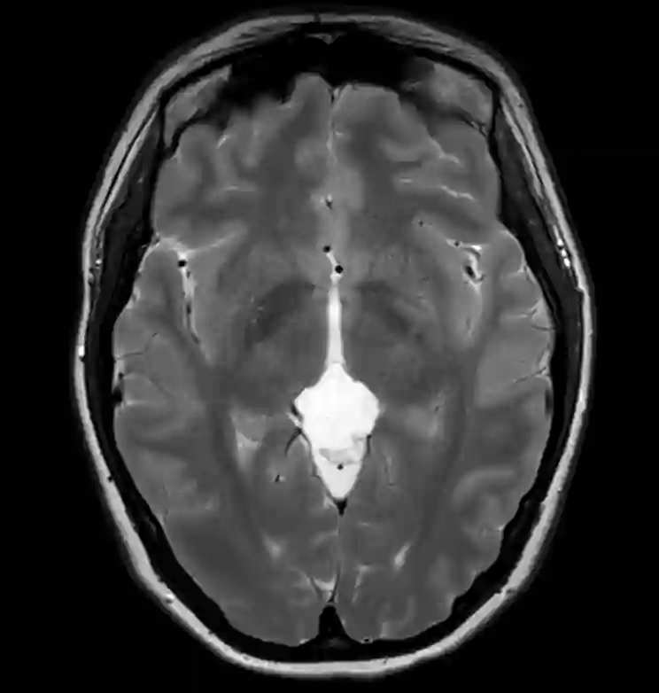
MRI
The gold standard for diagnosis is MRI. In the native T1 sequence, signal intensity can vary depending on the protein content of the cyst fluid and appear isointense to slightly hyperintense compared to cerebrospinal fluid. In the T2 sequence, a hyperintensity is often seen. In the T1 sequence with contrast agent, an enhancement in the cyst wall of up to a maximum of 2 mm can be seen in some cases. Nodular irregularities with contrast agent uptake may indicate another space-occupying lesion[^2].


Asymptomatic Pineal Cysts
In asymptomatic pineal cysts with a diameter of less than 2 cm and a typical imaging appearance with a wall thickness ≤ 2 mm and without irregularities or nodular contrast agent uptake, it is assumed that they do not show growth[^2]. However, the natural course cannot be predicted with certainty. In adults, the risk of cyst growth is about 4% at 6 months follow-up[^3] and in children about 11% at 10 months follow-up[^4]. Based on these data, it should be considered to conduct an early follow-up imaging after a few months of initial diagnosis[^2] to rule out rapid growth and subsequently increase the follow-up control interval.
Symptomatic Pineal Cysts
Symptoms
Pineal cysts can present symptomatically with hydrocephalic symptoms such as headaches, nausea, vomiting, and lethargy[^6]. It may also lead to papilledema, Parinaud syndrome or visual impairments[^6] [^7].
Surgical Treatment
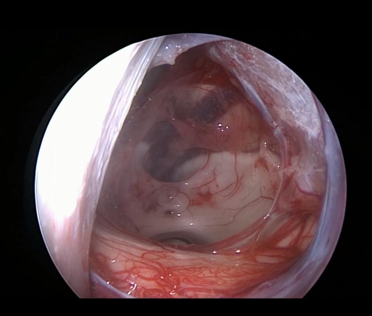
Surgical intervention is usually only necessary in extremely rare cases when the pineal cyst leads to an obstruction of cerebrospinal fluid flow and a consequent hydrocephalic buildup, or to symptoms such as headaches or visual disturbances[^8]. In symptomatic pineal cysts, surgical intervention is the therapy of choice[^2]. Pineal cysts can be resected, for example, via an infratentorial supracerebellar approach[^5] [^11].
-
pineal_cyst_resection.mp4 137.34 MB
- Video Beschreibung
- Operationsvideo einer mikrochirurgischen Resektion einer Pinealiszyste über einen infratentorielle supracerebellären Zugang.