Pedicle screw

Synonyms
Pedicle screw

Pedicle screws are used for posterior Stabilization of the Spine.

Structure

The screw head is either mobile (polyaxial), mounted on a ball-and-socket joint, or rigid (monoaxial), connected to the threaded shaft. The screw head contains a U-shaped recess that can accommodate a rod. This rod is secured in the screw head with a set screw. The pedicle screw can be used as an instrument to return a displaced vertebra to its proper position. To stabilize the spine, four or more pedicle screws are connected with rods along the spinal axis.

Carbon-fiber pedicle screws

Carbon-fiber pedicle screws have been available for several years. Carbon-fiber pedicle screws have the advantage of causing fewer artifacts on MRI imaging[^3]. This is particularly beneficial for patients with spinal tumors, in order to plan optimal follow-up treatment.

Pedicle screw entry point

Lumbar

In lumbar spondylodesis, the entry point of the pedicle screws is at the intersection of a line through the center of the Processus transversus and a line through the lateral portion of the superior Facet joint component[^1] [^2] [^4].

Schematic entry point for a pedicle screw
Lumbar pedicle screw entry point. At the thoracolumbar junction, pedicle screws are inserted at an angle of 5–10° to the midline. At L2, the screws are inserted at an angle of 10°, and at L5 at an angle of 15°, converging toward the midline. Figure adapted from Wallnoefer et al.

Imaging

Conventional pedicle screws cause artifacts on MRI, making MRI suboptimal for assessing pedicle screws. The gold standard for assessing the position of pedicle screws is CT imaging.
Pedicle screws on radiograph
Visualization of the pedicle screws of a multilevel spondylodesis on a lateral radiograph..

Complications

Pedicle screw breakage
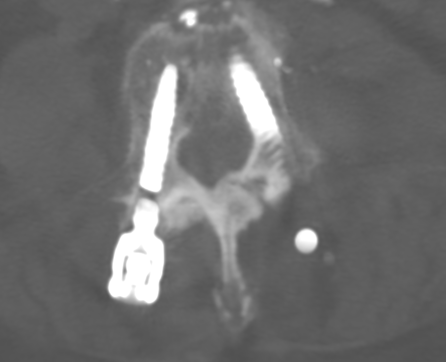
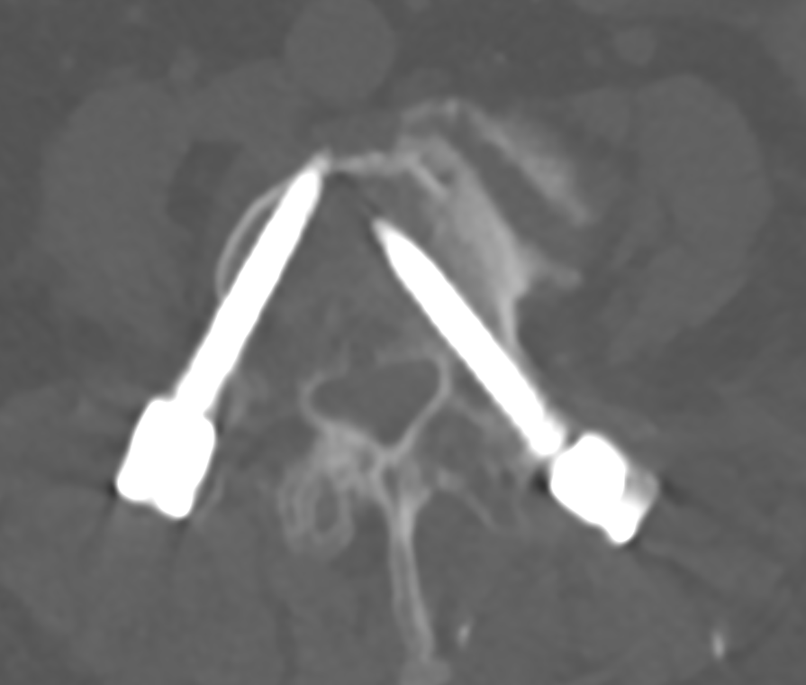
Broken pedicle screw on CT
Axial CT image of a right-sided broken pedicle screw.
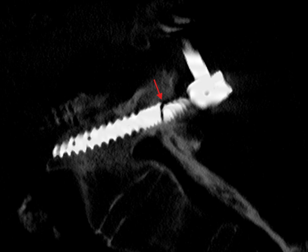
Fractured pedicle screw CT
Sagittal CT of a fractured pedicle screw (red arrow).
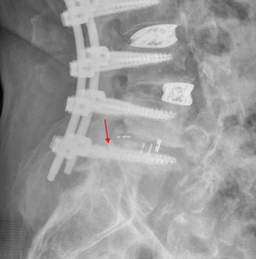
Fractured pedicle screw radiograph
Lateral radiograph of a fractured pedicle screw (red arrow).

Broken pedicle screws can, for example, be retrieved using a tap. A parallel surface can be created in the protruding portion of the pedicle screw with a tap, allowing the screw to then be unscrewed.
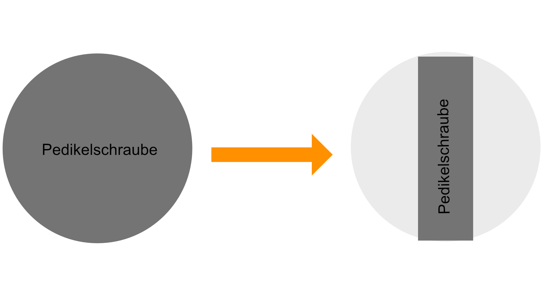
Retrieval technique for broken pedicle screws
Schematic illustration of a retrieval technique for broken pedicle screws without damaging the surrounding bone. A thread tap is used to create a parallel surface (the light gray portion is removed with the thread tap), allowing the pedicle screw to be unscrewed with a holding instrument.

Pedicle screw malposition

Malpositioned pedicle screw on CT
Axial CT showing a right-sided extrapedicular pedicle screw.

Pedicle screw migration

Pedicle screw migration refers to the “migration” of the pedicle screw from its original position.
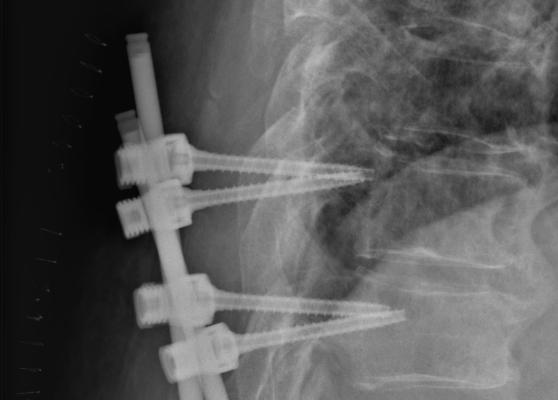
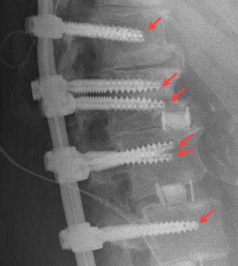
Pedicle screw migration on radiographs
Section of a lateral radiograph of a patient with migration of the pedicle screws at the two levels shown.

Pedicle screw loosening

Depending on its extent, loosening of the pedicle screws can be visualized on radiographs as well as on CT. This results in a hypodense rim around the pedicle screw.
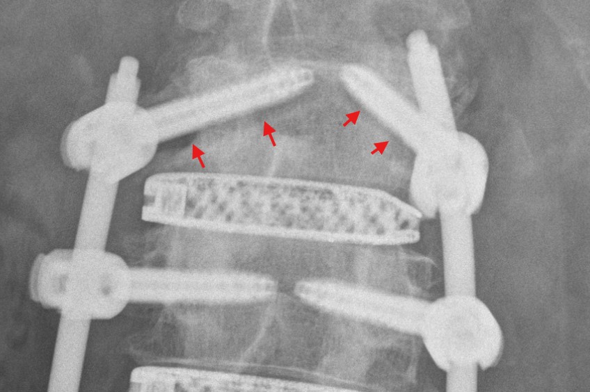
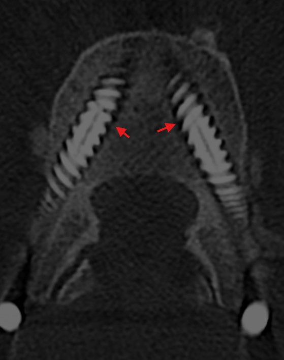
Radiograph of pedicle screw loosening
Radiograph showing bilateral radiolucent halos around lumbar pedicle screws.
CT halo sign around a pedicle screw
Axial CT image of a patient with bilateral loosening of lumbar pedicle screws. The red arrow indicates the radiolucent halo around the screw.

Editorial transparency

Authorship & revisions

Who created and updated this article, and which changes are documented.

Article created by
Last edited:
on
Revision history
6 edits · 1 contributor
Author list:
Show revision history 6 entries

6 edits by 1 author

Edit · Alaric Steinmetz · (+62 −31)

No content change recorded

Edit · Alaric Steinmetz ·

No content change recorded

Edit · Alaric Steinmetz · (+285 −0)

No content change recorded

Edit · Alaric Steinmetz · (+0 −281)

No content change recorded

Edit · Alaric Steinmetz ·

No content change recorded

Article created on · Alaric Steinmetz

Image viewer

Image

Open original