Choroid plexus papilloma is a rare benign brain tumor (WHO Grade I) that originates from the epithelium of the choroid plexus, the Choroid plexus.
Epidemiology
Choroid plexus papillomas can occur at any age, but approximately 70 % of patients are diagnosed before the age of 2 years[^1].
Location
Choroid plexus papillomas can occur in all areas where the choroid plexus is located. However, choroid plexus papillomas preferentially occur in the Lateral ventricle.
Imaging
CT imaging
On CT, most choroid plexus papillomas appear as an isodense/hyperdense lobulated mass with homogeneous contrast enhancement. Calcifications of varying severity occur in approximately 4 - 20 % of cases[^3].
MRI imaging
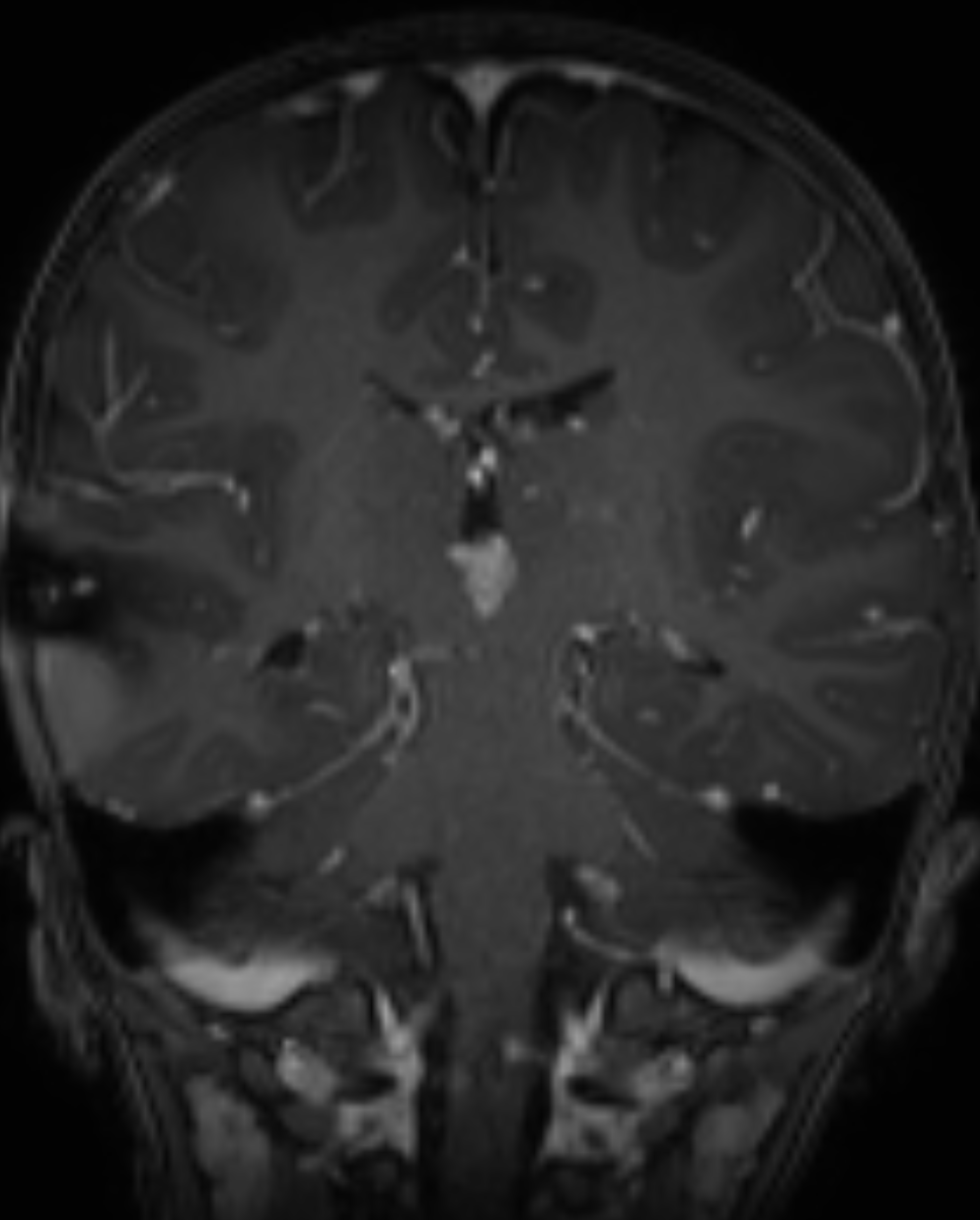
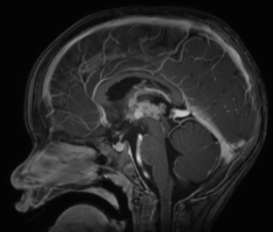
The gold standard for imaging choroid plexus papillomas is MRI imaging. On MRI, choroid plexus papillomas appear as a well-defined extra-axial lesion with fern-like projections, with contrast enhancement and no invasion of the brain parenchyma. In rare cases, choroid plexus papillomas may show little or no contrast enhancement[^3].
Prognosis
Malignant transformation may occur in rare cases; in a case series from 2007, malignant transformation into a WHO Grade 3 tumor within 59 months was observed in 2 of 124 patients[^2].