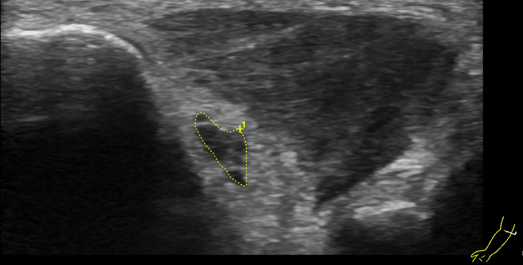
The ulnar nerve is a peripheral nerve arising from the medial cord of the brachial plexus. It contains motor and sensory nerve fibers originating from the C8 and Th1 nerve roots.
Course
The ulnar nerve branches off from the medial cord of the Brachial plexus. In the upper arm, the ulnar nerve gives off no branches and runs in the medial bicipital groove. At approximately the midpoint of the upper arm, it pierces the medial intermuscular septum of the arm and then passes dorsally around the medial epicondyle of the elbow into the Ulnar nerve groove, where it runs close to the surface. From here, the ulnar nerve travels together with the ulnar artery and vein beneath the flexor carpi ulnaris muscle toward the wrist. Here, the ulnar nerve does not pass through the Carpal tunnel, but rather through a fibrous canal, Guyon's canal, which extends over the flexor retinaculum and divides into its terminal branches, the sensory superficial branch and the motor deep branch. The ulnar nerve gives off smaller motor and sensory branches as early as its course through the forearm.

Function
Motor innervation
In the forearm, the ulnar nerve supplies the flexor carpi ulnaris muscle, which is responsible for flexion of the hand, as well as the ulnar half of the flexor digitorum profundus muscle, which is primarily responsible for flexion of the fourth and fifth fingers. Further motor branches arise from the deep branch, which forms an arch deep in the palmar aspect of the hand. It supplies the hypothenar muscles: the flexor digiti minimi brevis, abductor digiti minimi, and opponens digiti minimi muscles, which are responsible for flexion, abduction, and opposition of the little finger. It also provides motor innervation to all dorsal and palmar interosseous muscles, which are responsible for flexion at the metacarpophalangeal joints and extension at the interphalangeal joints. The deep branch also supplies the two ulnar lumbrical muscles. Finally, through its terminal motor branches in the thenar region, it innervates the adductor pollicis and the deep head of the flexor pollicis brevis.
Sensory innervation
Sensory innervation by the ulnar nerve covers the medial aspects of the dorsal and palmar surfaces of the hand; it supplies the entire little finger and only part of the adjacent fourth finger. At approximately the midpoint of the forearm, it gives off the dorsal branch, which supplies the medial dorsum of the hand and the dorsal skin of the little finger, as well as parts of the fourth finger and, at the base, also of the third finger. Somewhat farther distally, in the anterior third of the forearm, the palmar branch arises; it supplies the skin over the hypothenar eminence. The palmar skin of the little finger and the adjacent half of the fourth finger are innervated by the superficial branch, which arises at the level of the wrist during the terminal division of the nerve[^1].
Imaging
The ulnar nerve can be visualized using ultrasound or magnetic resonance imaging (MRI).