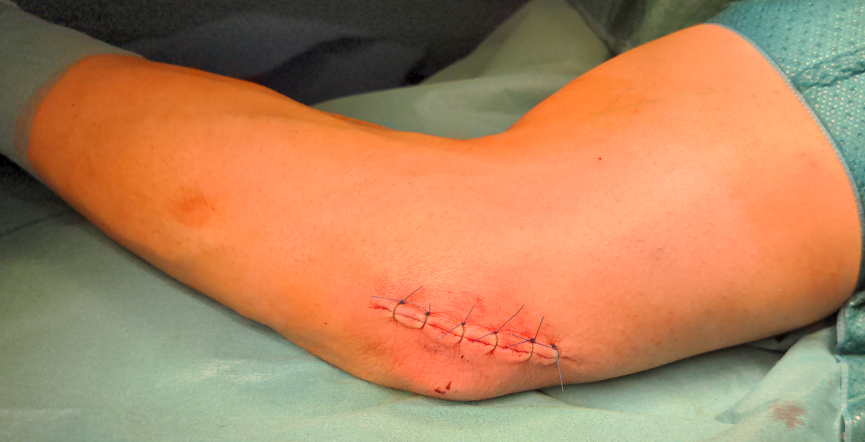
Cubital tunnel syndrome is a compression syndrome of the Ulnar nerve at the elbow.
Figure

Origin of the name
The term cubital tunnel syndrome has become established in the surgical literature. The name dates back to a publication by Feindel and Stratford in 1958, in which the term was used because of its similarity to carpal tunnel syndrome[^9] [^10].
Epidemiology
Cubital tunnel syndrome is the second most common compression syndrome of a peripheral nerve after Carpal tunnel syndrome. A study in the Italian population showed an incidence rate of 24.7 per 100,000 inhabitants[^1]. A study in the American population showed a similar incidence rate of 30.0 per 100,000 inhabitants[^2]. The incidence increases with increasing workload of the affected arm. Cubital tunnel syndrome occurs bilaterally in up to 40% of cases, with the left side affected almost three times as often as the right side, and men affected twice as often as women.
Symptoms
Typically, patients with cubital tunnel syndrome complain of numbness in the ulnar one and a half digits and along the ulnar border of the hand, as well as paresthesias in the distribution of the ulnar nerve. These occur particularly at night when the patient sleeps with the flexed arm under the head. Such symptoms are also frequently reported during activities involving a flexed arm or pressure on the elbow. Patients also report pulling pain at the elbow and along the ulnar forearm. In more advanced stages, motor impairment of the hand occurs, resulting from paresis of the interossei muscles and the adductor pollicis muscle[^11].
Neurological examination
On neurological examination, patients with cubital tunnel syndrome may have a positive Froment sign and Hoffmann-Tinel sign.
Inspection may reveal atrophy of the interossei muscles, particularly the first dorsal interosseous muscle, as well as the hypothenar muscles. In advanced stages, clawing of the fourth and fifth fingers may occur.
Electrophysiology
EMG
When cubital tunnel syndrome progresses to demyelination, EMG examination shows abnormal spontaneous activity and reduced motor unit recruitment[^3].
In more severe cases of cubital tunnel syndrome or with prolonged ulnar compression, axonal loss occurs. At this stage, electromyography shows abnormal insertional activity, indicating muscle denervation, while loss of motor axons leads to fibrillations at rest. The presence of motor unit action potentials during the recruitment phase indicates attempted reinnervation, either through collateral sprouting or axonal reinnervation[^4].
NCS
Additional nerve conduction findings are helpful in confirming the location and determining the severity of ulnar nerve compression. According to the summary statement of the »American Association of Neuromuscular and Electrodiagnostic Medicine«, the strongest evidence for the diagnosis of cubital tunnel syndrome is the presence of the following nerve conduction velocities[^5]:
Segment | Nerve conduction velocity |
Across the elbow | Less than 50 m\/s |
Above the elbow compared with the segment below the elbow to the wrist | More than 10 m\/s |
Ultrasound
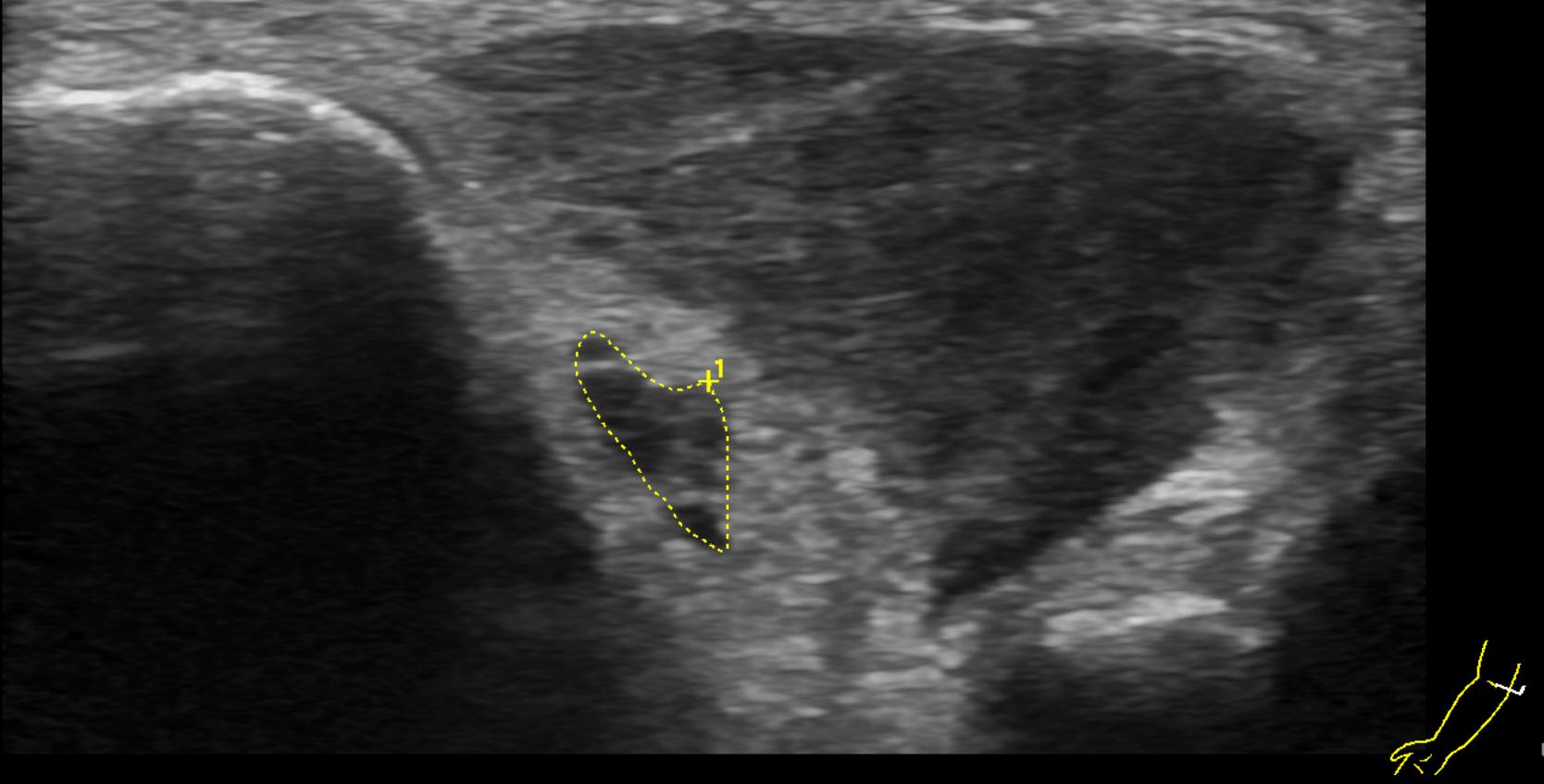
High-resolution ultrasound examination has become increasingly effective and specific for diagnosing cubital tunnel syndrome in recent years. In a study of patients with cubital tunnel syndrome but normal electrophysiological EMNG findings, ultrasound showed an enlarged cross-sectional area of the ulnar nerve near the elbow[^6].
A similar study by Volpe et al. from 2009[^7] showed that the maximum cross-sectional area, defined by enlargement of the ulnar nerve near the elbow, was 14.6 mm² in patients with confirmed ulnar neuropathy at the elbow compared with 7.1 mm² in control patients. Patients with axonal loss had a mean cross-sectional area of 18.3 mm², whereas patients with less severe symptoms, such as demyelination, had a cross-sectional area of 11.1 mm².
MRI
MRI imaging is helpful for evaluating soft-tissue structures and characterizing the extent and localization of the lesion. In one study, MRI was compared with EMG examinations, and MRI was found to be 25% more sensitive than EMG for diagnosing cubital tunnel syndrome. The most common MRI findings included a combination of increased signal intensity and nerve enlargement (63%), followed by nerve compression (27%), isolated increased signal intensity (23%), and isolated nerve enlargement (2%) [^8].

Treatment
Cubital tunnel syndrome can be treated conservatively or surgically, depending on its severity.