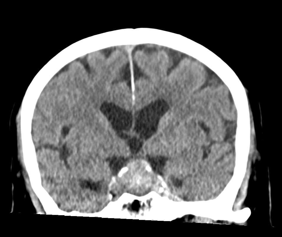
Pituitary adenomas are adenomas, a group of benign tumors, and arise from the tissue of the adenohypophysis. Surgical removal of the tumor is not necessary in all cases. Pituitary adenomas are classified as macroadenomas or microadenomas according to their size[^2].
Classification
Size | Designation |
< 3 mm | Pituitary picoadenoma[^3] |
< 10 mm | Pituitary microadenoma |
~ 10 mm | |
≥ 10 mm | Pituitary macroadenoma |
Hormone-secreting pituitary adenomas
Approximately 65 % of pituitary adenomas are hormone-secreting adenomas, with 48 % producing prolactin, 10 % growth hormone, 6 % ACTH, and 1 % TSH[^1]
Mass effect caused by pituitary macroadenomas
The following structures are frequently affected by the mass effect of pituitary macroadenomas:
optic chiasm: This may result in bitemporal hemianopia and decreased visual acuity.
Third ventricle: Compression of the third ventricle can result in obstructive hydrocephalus.
Cavernous sinus: This can result in deficits of the cranial nerves that run through the cavernous sinus.
Imaging
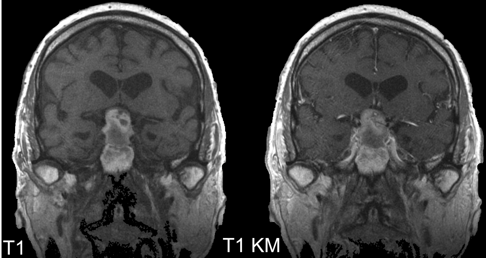
MRI
The gold standard for the imaging diagnosis of pituitary adenomas is MRI imaging.


CT
Depending on their size, pituitary adenomas can also be visualized on CT imaging.