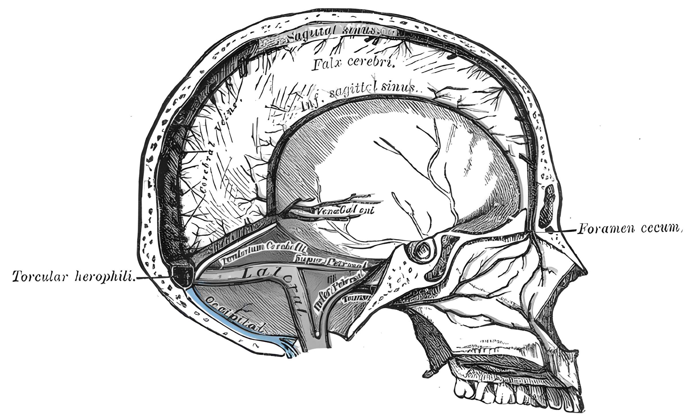
The occipital sinus is a highly variable intradural cerebral venous sinus that connects the Marginal sinus with the Confluence of sinuses.
Epidemiology
In a cadaver study of 200 patients, a distinct occipital sinus could be identified in 64,5% of cases[^1].
Anatomy
The occipital sinus begins in several small channels at the margin of the Foramen magnum and courses between the cerebellar hemispheres, embedded in the Falx cerebelli. In a cadaver study of 200 specimens, five different morphological variants could be distinguished[^1]:
A single occipital sinus in the midline of the posterior cranial fossa could be identified in 38% of cases. In 18% of these cases, the occipital sinus divided into two venous channels anterior to the posterior margin of the Foramen magnum.
A double occipital sinus was observed in 22,% of cases.
A single left-sided occipital sinus was observed in 4% of cases.
A single right-sided occipital sinus was observed in 3% of cases.
No occipital sinus could be identified in 35,5% of cases.
Clinical relevance
Surgical occlusion of the occipital sinus is generally without consequences. Surgical opening of a well-developed occipital sinus can result in severe venous bleeding intraoperatively, so preoperative identification of an occipital sinus on imaging may be helpful for surgical procedures in the region of the posterior cranial fossa.