An abscess is a collection of pus in a non-preformed body cavity caused by inflammatory tissue liquefaction. A symptomatic intracranial or spinal abscess is, in most cases, an urgent indication for surgical removal.
Location
A neurosurgically relevant abscess may occur intraspinally or intracerebrally.
Imaging
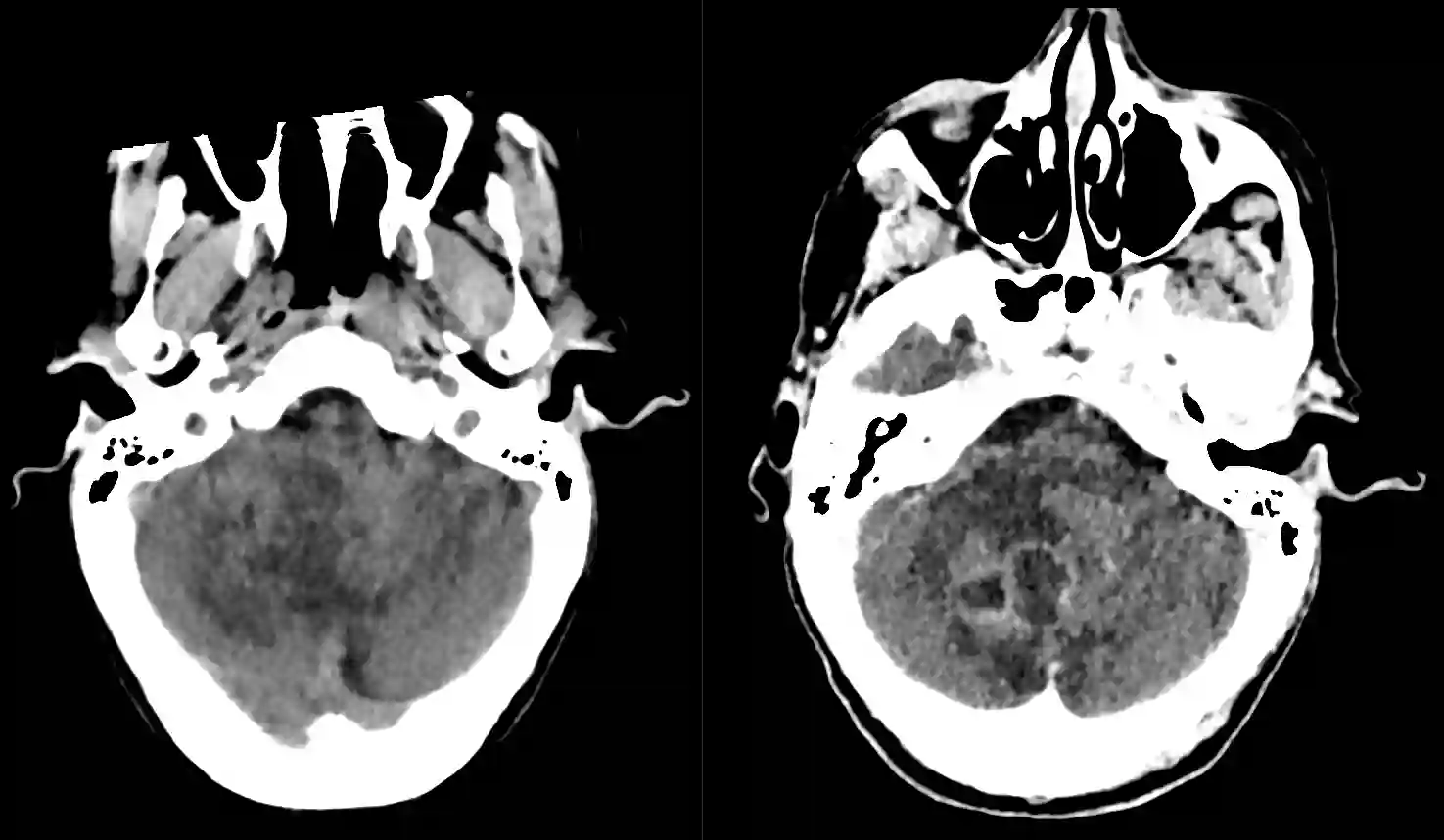
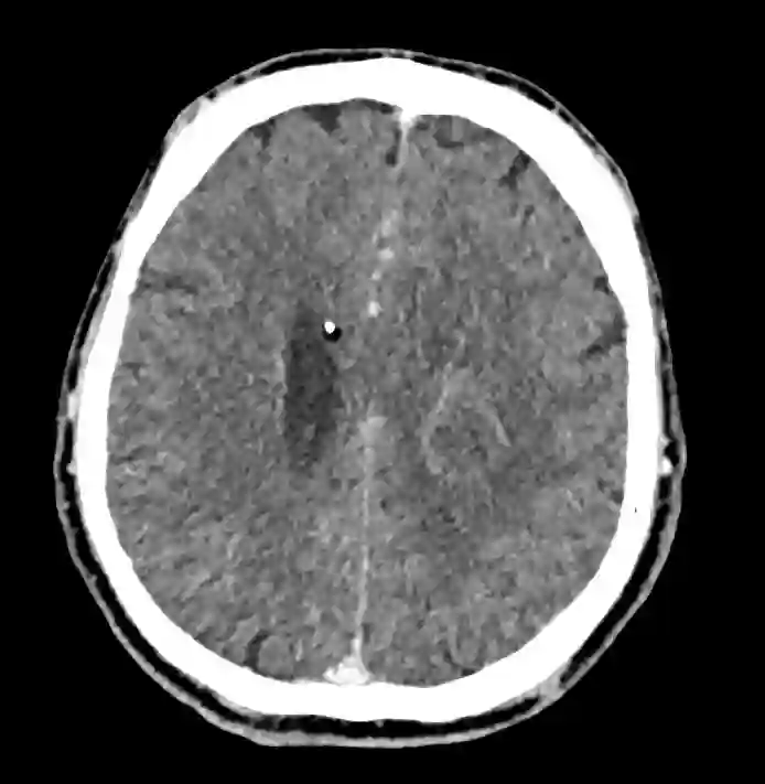
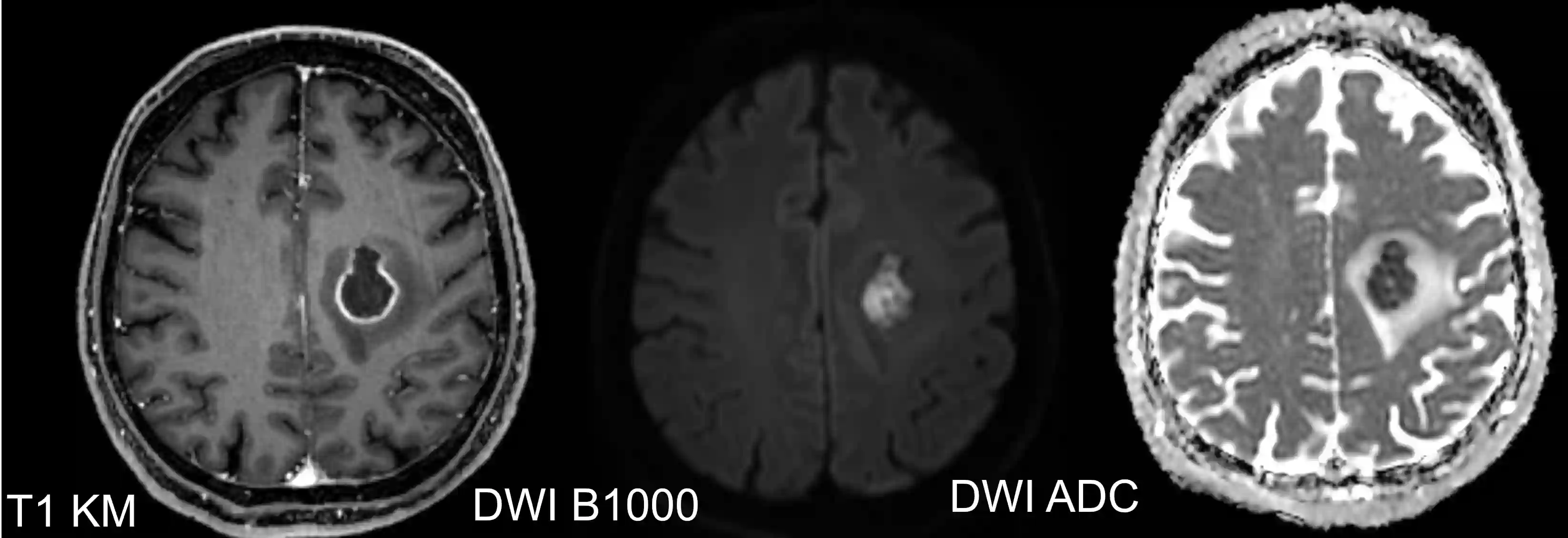
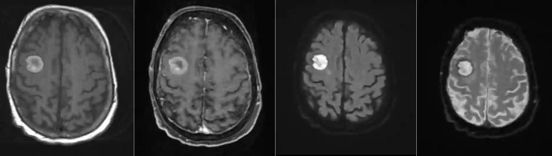
For diagnostic purposes, intracerebral abscesses show the following characteristics on MRI imaging[^1]:
MRI T1 with contrast medium | MRI ADC sequence | ||
Ring enhancement of the lesion with contrast medium | Subtle ring enhancement at the edge of the lesion | Hyperintense | Hypointense |
Computed tomography


\n MRI


\n Treatment
Patients with an intracranial or spinal abscess require antibiotic therapy for several weeks even after surgical treatment; the regimen depends on the pathogen identified microbiologically. The occurrence of intracranial abscesses correlates with patients' socioeconomic status and therefore represents a major challenge in developing countries[^2].
Pathology
Histologically, brain abscesses can be divided into four different stages and differ in resistance during surgical puncture and aspiration[^1]. Progression through the various stages takes at least two weeks and is prolonged by administration of Steroids[^1].
Stage | Designation | Time period | Histological characteristics | Resistance during aspiration |
1 | Early Cerebritis | Day 1-3 | Early infectious and inflammatory phase. Poor demarcation from the surrounding brain tissue. Toxic changes in neurons and perivascular infiltrates. | Intermediate resistance |
2 | Late cerebritis | Day 4-9 | Reticular matrix and development of a necrotic center | No resistance |
3 | Early capsule formation | Day 10-13 | Neovascularization and central necrosis with surrounding reticular network. | No resistance |
4 | Late capsule | > Day 14 | Collagen capsule, necrotic center, and gliosis surrounding the capsule | Strong resistance and a "pop" during capsule puncture |
Microbiology
Brain abscesses often consist of a mixed flora. Microaerophilic and anaerobic streptococci are the most frequently identified pathogens[^3].