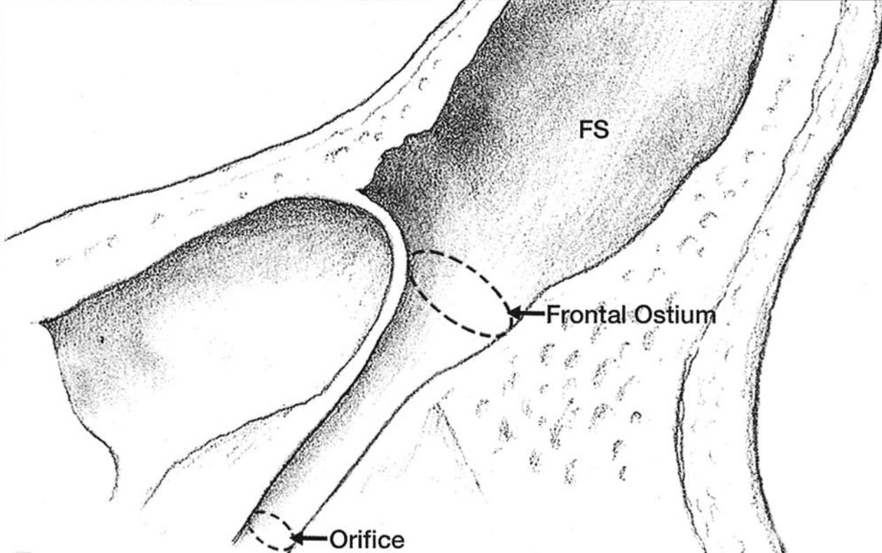
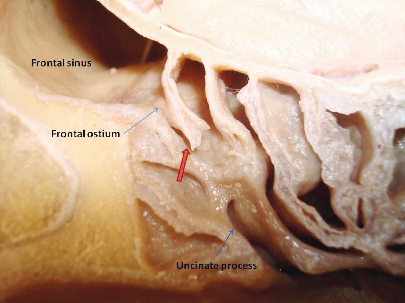
The ductus frontonasalis connects the Frontal sinus to the middle nasal meatus[^1].
Anatomy
The ductus frontonasalis is lined with respiratory epithelium.
Figure
EnlargeSchematic illustration of the ductus frontonasalis. FS = sinus frontalis. Figure adapted from Kim, Kyung Su, et al. Surgical anatomy of the nasofrontal duct: anatomical and computed tomographic analysis. The Laryngoscope 111.4 (2001): 603-608.EnlargeVisualization of the ductus frontonasalis (red arrow) in an anatomical specimen. Figure adapted from Gupta, Tulika, Anjali Aggarwal, and Daisy Sahni. Surgical anatomy of the frontal sinus outflow pathway: a cadaveric study. Eur J Anat 17.1 (2013) 29-34.
Imaging
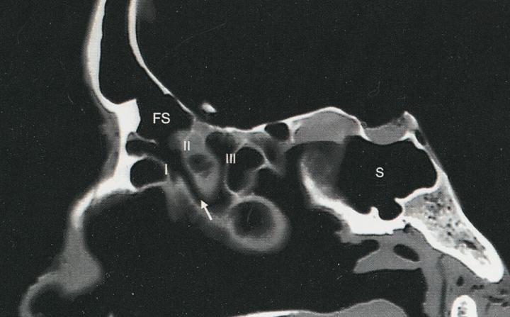
The ductus frontonasalis can be visualized on CT imaging.
EnlargeSagittal CT imaging showing the ductus frontonasalis (white arrow). FS = sinus frontalis. Figure adapted from Kim, Kyung Su, et al. "Surgical anatomy of the nasofrontal duct: anatomical and computed tomographic analysis." The Laryngoscope 111.4 (2001): 603-608.
Clinical relevance
Obstruction of the ductus frontonasalis, for example by polyps or tumors, can lead to frontal sinusitis [^2].
When the frontal sinus is cranialized, the mucosa of the frontal sinus should be removed and the frontal sinus obliterated with fat or muscle to prevent communication with the outside through the ductus frontonasalis[^3].
Weekly update
Newsletter
Relevant neurosurgical studies, concisely summarized in a weekly email newsletter.
By subscribing, you consent to receiving the newsletter through Buttondown. Unsubscribe using the link in any email.
Privacy policy