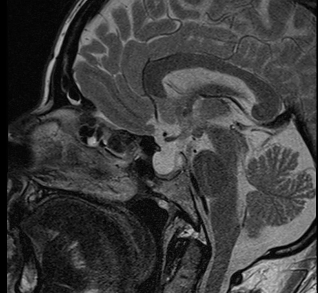
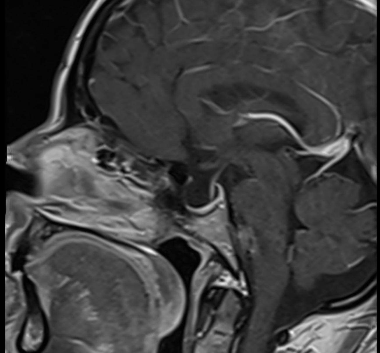
Empty sella syndrome is classified as either primary or secondary empty sella syndrome.
Primary Empty Sella Syndrome
Primary empty sella syndrome is characterized by herniation of the arachnoid into the Sella turcica, where CSF pulsations[^1] probably cause a mass effect, consequently resulting in radiographic enlargement of the sella turcica and displacement of the Pituitary gland to the floor of the sella turcica. Primary empty sella syndrome is more common in women and is associated with obesity and arterial hypertension. The most common symptom of primary empty sella syndrome is headache, which may also occur together with Cerebrospinal fluid rhinorrhea. Surgical repair may be necessary in patients with CSF rhinorrhea; otherwise, surgical treatment is usually not indicated[^2].
Secondary Empty Sella Syndrome
Secondary empty sella syndrome may occur after trauma [^3] or after a surgical procedure involving the sella turcica. It may also occur in conditions associated with increased intracranial pressure, such as idiopathic intracranial hypertension.

Imaging
The optimal imaging modality for evaluating empty sella syndrome is MRI imaging.