Chordomas are rare tumors that occur particularly in the region of the Clivus, but can also occur in the region of the Spine[^1]. Chordomas do not arise from bone tissue, but from remnants of the notochord.
Epidemiology
Chordomas are rare tumors with an incidence of approximately 0.08/100,000 per year[^4] and account for approximately 1-4 % of all bone malignancies[^1]. Chordomas can occur at any age, but are particularly common in patients between the 3rd and 6th decades of life[^1].
Imaging
The gold standard for imaging chordomas is MRI imaging.
MRI
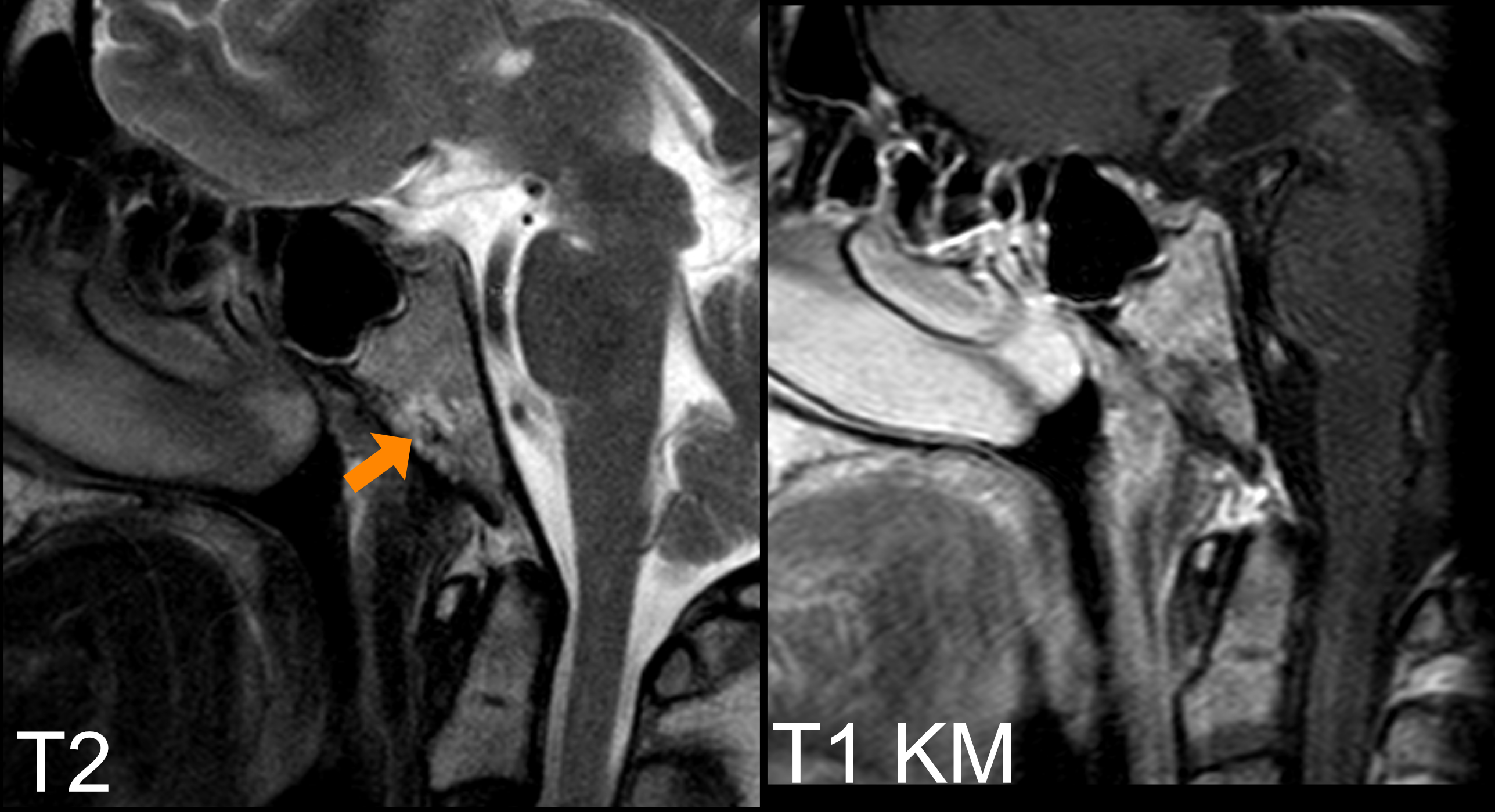
On MRI, chordomas typically appear as follows[^3]:
Noncontrast T1 | Contrast-enhanced T1 | T2 |
Iso- to Hypointense | Heterogeneous contrast enhancement |

CT
Clival chordomas in particular frequently show osseous destruction of the clivus and calcifications[^3].
Histopathology
Chordomas can be classified into three different histopathological types[^3]:
Chordoma, not otherwise specified (NOS)
Chondroid chordoma
Dedifferentiated chordoma

Immunohistochemistry
The presence of physaliphorous cells and positive immunohistochemical staining for Brachyury are typical[^6].
Treatment
The treatment of choice for clival chordomas is surgical resection followed by proton beam radiotherapy, as radiotherapy usually requires high doses and clival chordomas are located close to critical structures. Chordomas are chemoresistant tumors[^3].
Prognosis
Patients with a newly diagnosed chordoma have a median survival time of 6.29 years, with the following survival rates[^1]:
Time period | Survival rate |
5 years | 47 % - 80 % |
10 years | 40 % |
20 years | 13 % |