Astroblastoma is an extremely rare[^1] GFAP-positive glial tumor that occurs in children and young adults[^2].
Historical background
Astroblastoma was first described in 1930 by Bailey and Cushing[^5].
Epidemiology
Astroblastomas are rare brain tumors and account for approximately 0.45% to 2,% of all gliomas[^3] [^4].
Imaging
On imaging, astroblastomas frequently appear as well-demarcated tumors with solid and cystic components. Astroblastomas frequently demonstrate intratumoral cysts and a "bubbly" appearance [^6].
MRI
On MRI imaging, astroblastomas frequently appear hyperintense relative to white matter on T2 and FLAIR sequences and hypo- to isointense on T1 sequences. The tumor typically demonstrates heterogeneous contrast enhancement.
CT
On CT imaging, astroblastomas, as on MRI, demonstrate heterogeneous contrast enhancement. Calcifications are also frequently seen on CT [^6].
Histopathology
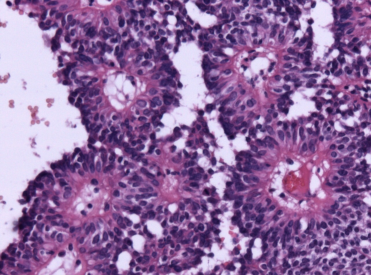
The tumor is characterized by astroblastic pseudorosettes, which are formed by tumor tissue tapering toward central blood vessels[^2].