Arteriovenous malformations are convolutions of pathological vessels that form a shunt connection between an artery and a vein and therefore do not form a capillary bed to supply the neural parenchyma. In combination with the still relatively high arterial pressure and venous congestion in the vessels, this makes AVMs highly susceptible to hemorrhage.
Diagnostics
Digital subtraction angiography
Digital Digital subtraction angiography (DSA) serves as the gold standard for the diagnosis of AVMs.

MRI
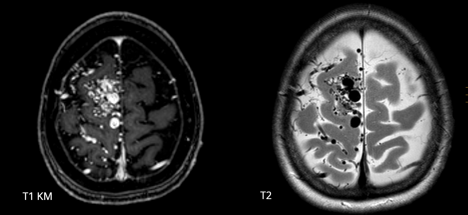
MRI imaging plays a subordinate role in the diagnosis of arteriovenous malformations. The initial diagnosis is often made through MRI imaging. However, digital subtraction angiography remains the gold standard for precise diagnosis and treatment.
Classification
AVMs are generally classified according to the Spetzler–Martin classification.
Hemorrhage risk
The unselected risk of rupture of an arteriovenous malformation is approximately 2.3 % per year[^1].
Treatment
Each arteriovenous malformation has unique anatomy and requires a tailored treatment approach. Treatment options range from endovascular procedures and microsurgical procedures to radiosurgical options.
Surgical treatment
AVMs corresponding to Spetzler grades 1 and 2 are best suited for surgical treatment alone[^5].
Treatment with stereotactic radiosurgery
Small-volume arteriovenous malformations that are located deep or in eloquent regions, or that occur in patients with a high operative risk due to comorbidities, can be treated with stereotactic radiosurgery[^2]. Radiosurgery causes endothelial damage, followed by proliferation of smooth muscle and thickening of the vessel wall, until the AVM becomes occluded. This process leading to occlusion takes approximately 1–3 years[^3]. During this period, the annual risk of hemorrhage remains approximately 1–3 %, corresponding to the natural risk of rupture without treatment[^4].
Videos
Angiography of a Spetzler-Martin grade 2 AVM in the frontal lobe
Angiography of an arteriovenous malformation (Martin-Spetzler grade 2) in the frontal lobe, supplied primarily by the middle cerebral artery and draining via a cortical vein into the superior sagittal sinus.