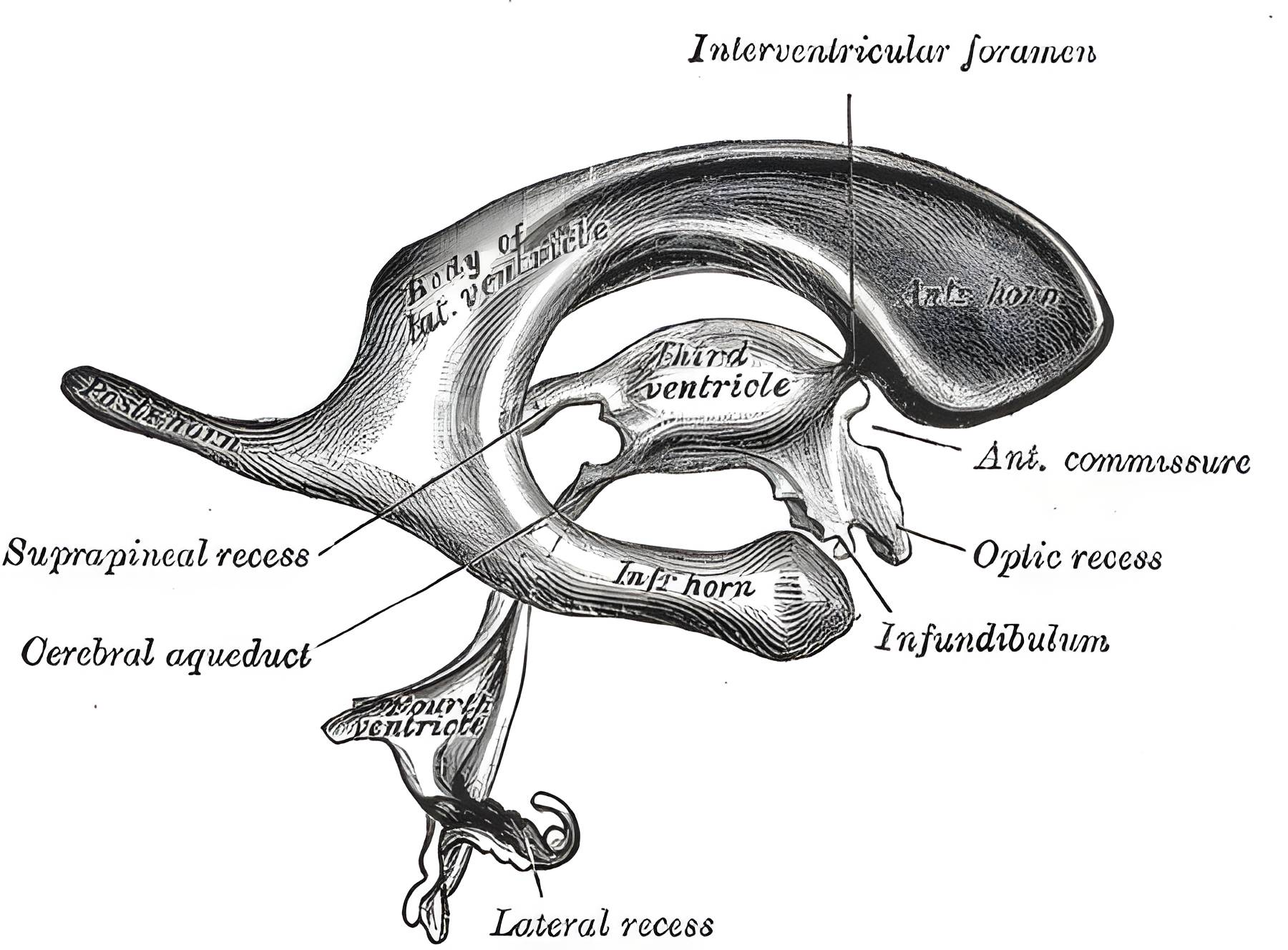
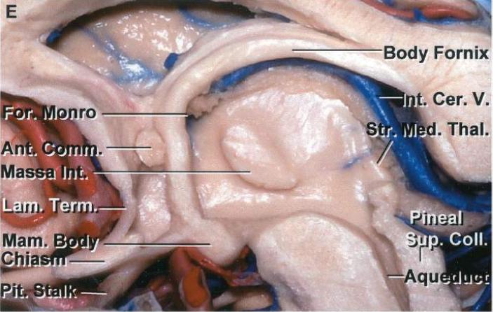
The Aquaeductus mesencephali is the connecting channel between the third- and the fourth ventricle.
Anatomy
The Aquaeductus mesencephali runs medially along the posterior surface of the Midbrain to the rhomboid fossa, where it expands into the fourth ventricle. Unlike the ventricles, it is not covered by portions of the Choroid plexus. The Periaqueductal gray matter surrounds the aqueduct circumferentially.
Figure
EnlargeSchematic illustration of the ventricular system and its components. Figure adapted from Gray’s Anatomy, Figure 736.EnlargeSagittal section through the third ventricle. Figure adapted from Albert Rhoton 2002 in Neurosurgery.
Imaging
EnlargeVisualization of the aquaeductus mesencephali on a sagittal MRI image (red arrow).
Weekly update
Newsletter
Relevant neurosurgical studies, concisely summarized in a weekly email newsletter.
By subscribing, you consent to receiving the newsletter through Buttondown. Unsubscribe using the link in any email.
Privacy policy