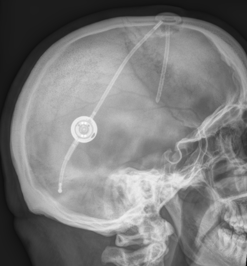
A ventriculoperitoneal shunt (VP shunt) is a mechanical connection that enables the drainage of cerebrospinal fluid from the brain into the abdomen. VP shunts usually have valves that, depending on the model, allow the opening pressure to be adjusted externally. In most cases, the valve setting is changed using a magnetic mechanism. The malfunction rate of VP shunts is approximately 40% after two years, meaning that approximately 60% of VP shunts are still functioning after 2 years1. The risk of a postoperative infection in the VP shunt system within the first year is 4-10%2.
Gravitational unit
A gravitational unit can be installed in addition to a conventional shunt valve. The gravitational unit can minimize the risk of overdrainage because it introduces additional resistance into the shunt system when the patient is standing. The gravitational unit is active only in the vertical position and inactive in the horizontal position.
Complications
In addition to general complications such as shunt overdrainage or underdrainage, placement of the peritoneal catheter carries specific associated risks:
- VP shunt-associated ascites
- Volvulus3
- Inguinal hernias4
- Peritonitis
- Hydrocele
- Obstruction of the abdominal catheter tip
- Migration of the abdominal catheter tip, for example into the scrotum5

References
Book: Neurosurgical Knowledge Update - A comprehensive review. Robert E. Harbaugh et al. Thieme Verlag ↩ ↩ ↩ ↩
Sakoda, Thomas H., John A. Maxwell, and Charles E. Brackett. "Intestinal volvulus secondary to a ventriculoperitoneal shunt: Case report." Journal of neurosurgery 35.1 (1971): 95-96. ↩ ↩
Moazam, F., et al. "Inguinal hernias after ventriculoperitoneal shunt procedures in pediatric patients." Surgery, gynecology & obstetrics 159.6 (1984): 570-572. ↩ ↩
Ram, Zvi, et al. "Ventriculoperitoneal shunt malfunction due to migration of the abdominal catheter into the scrotum." Journal of pediatric surgery 22.11 (1987): 1045-1046. ↩ ↩