Pneumocephalus refers to the presence of gas, usually air, within the neurocranium.
Causes
Pneumocephalus may occur, for example, after intracranial surgery, in the setting of an open traumatic brain injury, or with an intracranial infection.
Imaging
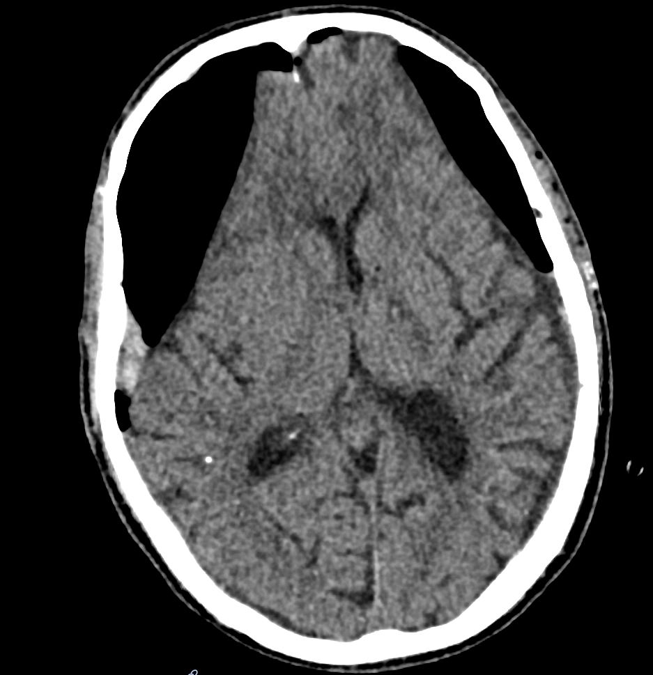
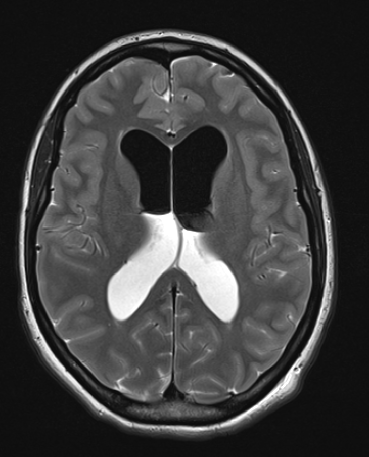
Intracranial air collections are best visualized on CT imaging and MRI imaging. Marked pneumocephalus with compression of the frontal lobe may appear on CT imaging as the so-called Mount Fuji sign.
CT


MRI
Treatment of postoperative pneumocephalus
Marked or symptomatic postoperative pneumocephalus can be treated by increasing the FiO2. Increasing the FiO2 leads to enhanced resorption of intracranial air[^2]. Administration of 100 % FiO2 can be tolerated for 24-48 hours without severe pulmonary toxicity[^3]. Tension pneumocephalus can cause severe symptoms and should be evacuated with urgency similar to that for an intracranial hemorrhage[^1].