Pleomorphic Xanthoastrocytoma (WHO Grade 2-3)
Last edit by Alaric Steinmetz on
Synonyms: pleomorphic xanthoastrocytoma, PXA
Pleomorphic xanthoastrocytomas are extremely rare primary brain tumors that belong to the group of astrocytomas.
Epidemiology
Pleomorphic xanthoastrocytomas account for less than 1% of all astrocytomas. At most, the incidence is 0.51/1000000 between the ages of 10 and 30 years1.
WHO Classification
According to the 2021 WHO Classification of Tumors of the Central Nervous System, pleomorphic xanthoastrocytomas are classified as WHO grade 2 and 32.
Localization
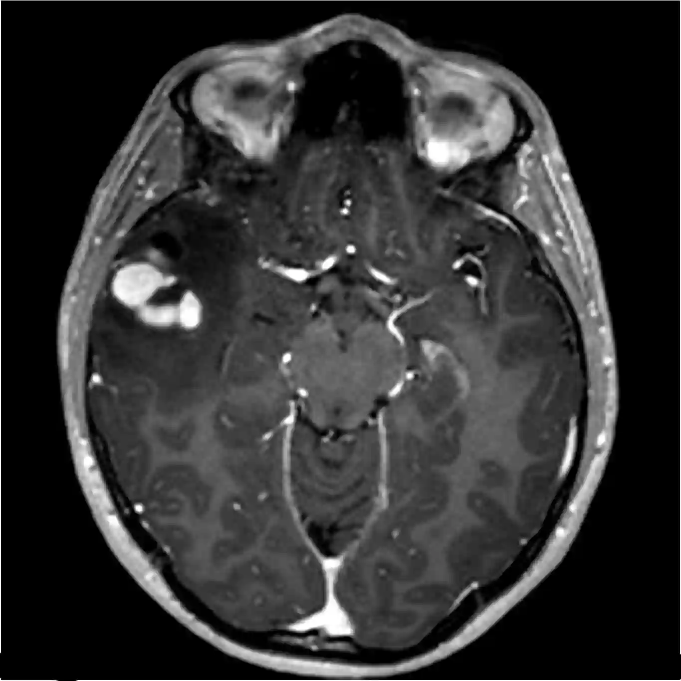
Pleomorphic xanthoastrocytomas frequently exhibit superficial growth along the gyri, preferentially in the temporal lobe, with contact with and infiltration of the leptomeninges. More than 90% of pleomorphic xanthoastrocytomas are located supratentorially, and in > 67% of cases they have contact with the dura3.
Symptoms
In accordance with their cortical localization, pleomorphic xanthoastrocytomas frequently become symptomatic through epileptic seizures.
Imaging
On MRI, pleomorphic xanthoastrocytomas appear as well-demarcated, sometimes cystic tumors that resemble pilocytic astrocytoma. Accordingly, in addition to cysts, there are vital nodules that enhance with contrast material4. Pleomorphic xanthoastrocytomas frequently involve the dura mater5.

References
Lettau, M. "Pleomorphic xanthoastrocytomas–Pleomorphic xanthoastrocytomas." RöFo-Fortschritte auf dem Gebiet der Röntgenstrahlen und der bildgebenden Verfahren. Vol. 187. No. 01. Georg Thieme Verlag KG, 2015. ↩ ↩
Louis, David N., et al. "The 2021 WHO classification of tumors of the central nervous system: a summary." Neuro-oncology 23.8 (2021): 1231-1251. ↩ ↩
Handbook of Neurosurgery. Greenberg M, ed. 9th Edition. New York: Thieme; 2019. ↩ ↩
Forsting M, Uhlenbrock D, ed. MRI and MRA of the Head. 2nd, completely revised and expanded edition. Stuttgart: Thieme; 2011 ↩ ↩
Gaudino, S., Martucci, M., Russo, R. et al. MR imaging of brain pilocytic astrocytoma: beyond the stereotype of benign astrocytoma. Childs Nerv Syst 33, 35–54 (2017) ↩ ↩