Os sacrum

Synonyms
Kreuzbein
ICD-11
XA14W3

The os sacrum is a bone of the Spine formed by the five sacral vertebrae and the intervertebral discs between them.

Anatomy

The os sacrum lies between the two ossa coxae and articulates cranially with the fifth lumbar vertebra (L5) via the lumbosacral joint and caudally with the Coccyx. The os sacrum has an anteriorly concave surface, the facies pelvina, and a posteriorly oriented convex facies dorsalis. The surface facing the last true lumbar vertebra is referred to as the basis ossis sacri[^1].

Fractures

Sacral fractures may occur after high-energy trauma or as insufficiency fractures. Neurological deficits occur particularly when the sacral canal or sacral neuroforamina are involved. Fractures of the os sacrum can be classified according to the Gibbons classification.

Figure
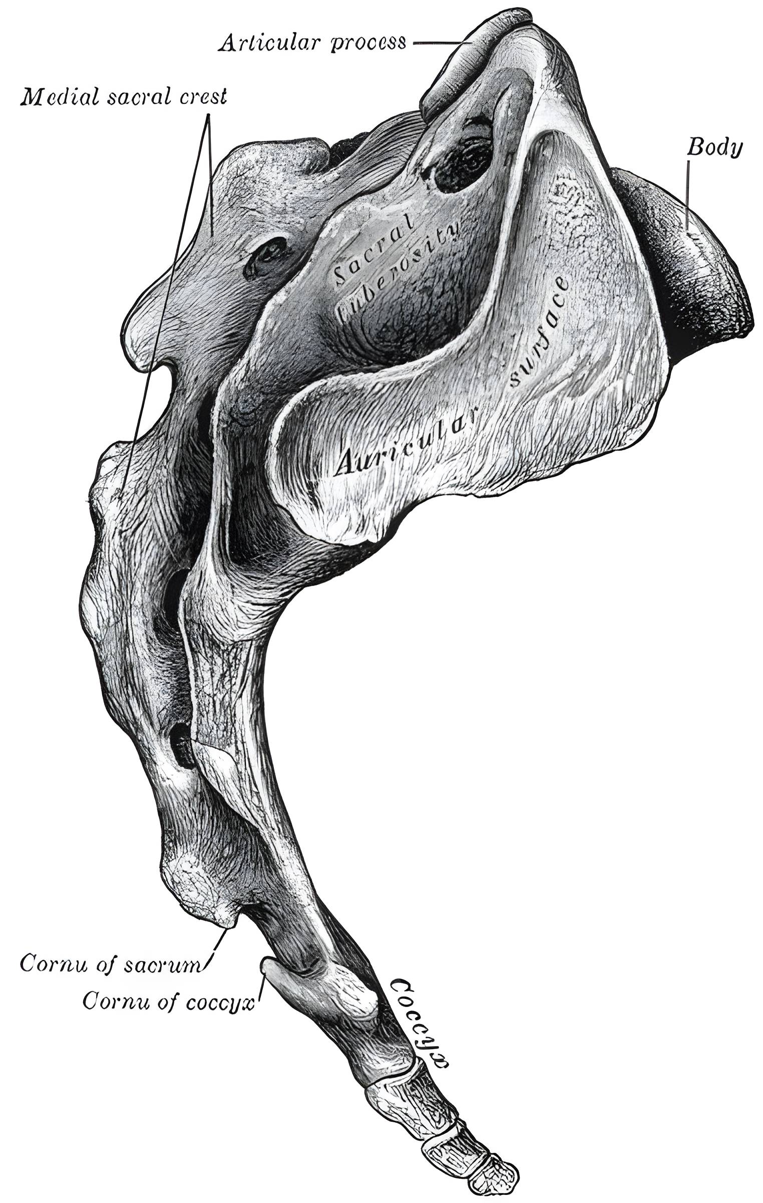
Sacrum, Figure 97
Schematic lateral illustration of the sacrum. Figure adapted from Gray's Anatomy, Figure 97.
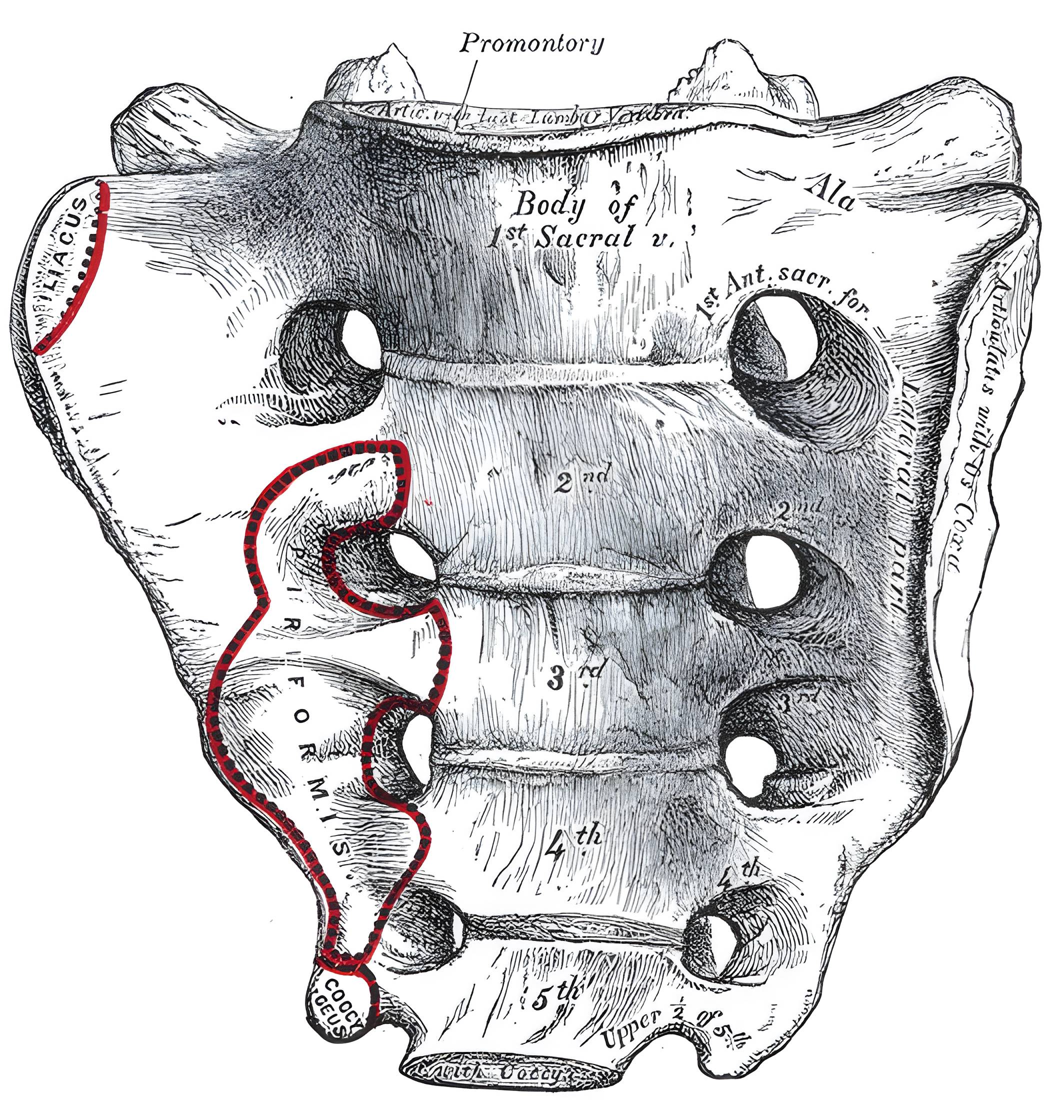
Sacrum, Figure 95
Schematic illustration of the sacrum. Figure adapted from Gray's Anatomy, Figure 95.
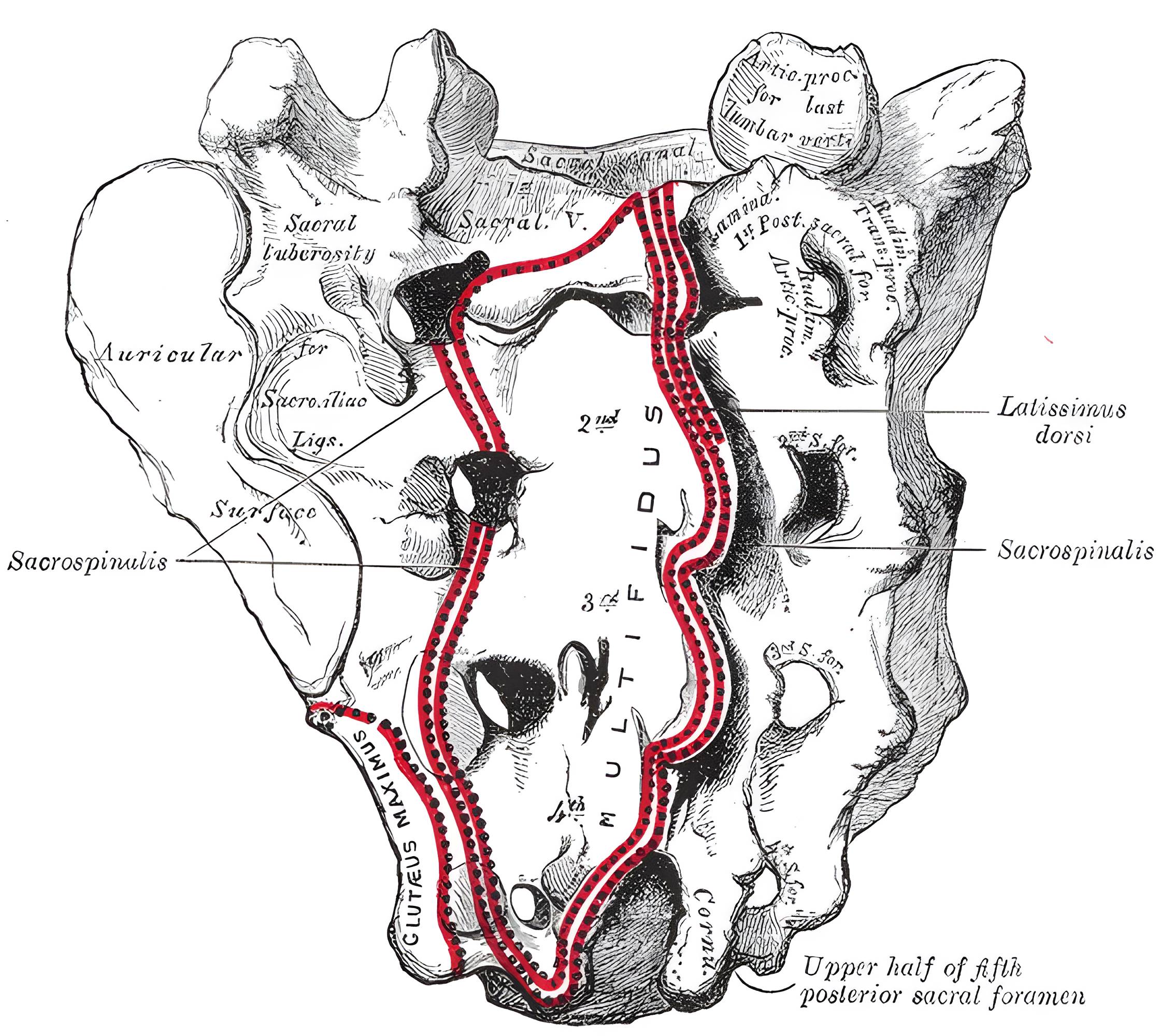
Sacrum, Figure 96
Schematic illustration of the sacrum. Figure adapted from Gray's Anatomy, Figure 96.

Imaging

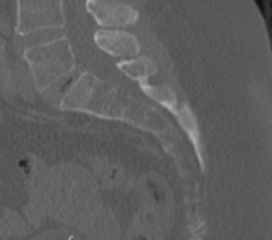
Sagittal CT image of the sacrum
Sagittal CT image of the sacrum.
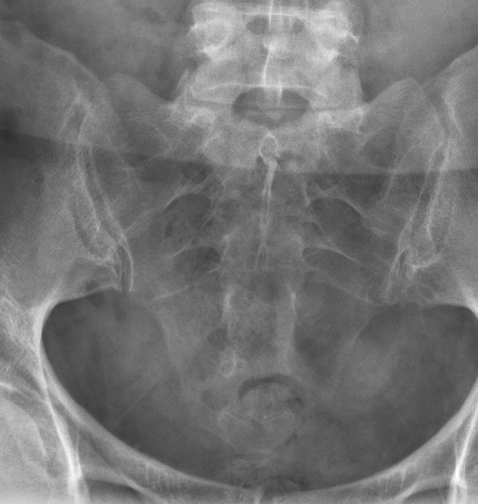
Os sacrum Röntgen
Darstellung des Os sacrum im a/p Röntgen.

Editorial transparency

Authorship & revisions

Who created and updated this article, and which changes are documented.

Article created by
Last edited:
on
Revision history
8 edits · 1 contributor
Author list:
Show revision history 8 entries

8 edits by 1 author

Edit · Alaric Steinmetz · (+124 −29)
  • Text added: “Anatomy”
  • Text added: “The os sacrum lies between the two ossa coxae and articulates cranially with the fifth lumbar vertebra (L5) via the lumbosacral joint and caudally with the Coccyx . The os sacrum has an anteriorly concave surface, the facies pelvina, and a posteriorly oriented convex facies dorsalis. The surface facing the last true lumbar vertebra is referred to as the basis ossis sacri[^1].”
  • Text added: “Fractures”
  • Text added: “Sacral fractures may occur after high-energy trauma or as insufficiency fractures. Neurological deficits occur particularly when the sacral canal or sacral neuroforamina are involved. Fractures of the os sacrum can be classified according to the Gibbons classification .”
  • Text added: “Figure”
  • Text added: “Imaging”
  • Text removed: “## Anatomy {#anatomie} The Os sacrum has an anteriorly concave surface, the Facies pelvina, and a posteriorly directed convex Facies dorsalis. The surface facing the last true lumbar vertebra is referred to as the basis ossis sacri[^1].”
  • Text removed: “## Fractures {#frakturen} Fractures of the Os sacrum can be classified according to the [Gibbons classification](\/scores-und-klassifikationen\/sakrumfraktur-klassifikation).”
  • Text removed: “## Figure {#abbildung}”
  • Text removed: “## Imaging {#bildgebung}”
  • Text removed: “**References**”
  • Text removed: “[^1]: Platzer, Werner. Taschenatlas Anatomie. Vol. 1. Georg Thieme Verlag, 2018”
  • Text changed: “The Os sacrum is a bone of the vertebral column formed from the 5 sacral vertebrae and the intervertebral discs between them.”“The os sacrum is a bone of the Spine formed by the five sacral vertebrae and the intervertebral discs between them.”
  • Citation removed: “Platzer, Werner. Taschenatlas Anatomie. Vol. 1. Georg Thieme Verlag, 2018”
  • Image added: os_sacrum_roentgen_ap.png
Edit · Alaric Steinmetz ·

No content change recorded

Edit · Alaric Steinmetz · (+44 −0)

No content change recorded

Edit · Alaric Steinmetz · (+0 −42)

No content change recorded

Edit · Alaric Steinmetz ·

No content change recorded

Edit · Alaric Steinmetz · (+44 −0)

No content change recorded

Edit · Alaric Steinmetz · (+0 −42)

No content change recorded

Article created on · Alaric Steinmetz

Image viewer

Image

Open original