The orbit refers to the bony eye socket. The orbit is approximately 4 to 5 cm deep and is shaped like a four-sided pyramid, with the apex posteriorly and the base at the entrance to the eye socket[^1].
Anatomy
Bones of the orbit
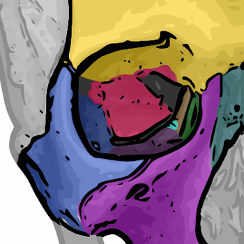
The orbit is formed by a total of 7 different bones:
Roof of the orbit
The roof of the orbit is formed anteriorly by the pars orbitalis ossis frontalis and posteriorly by the ala minor ossis sphenoidalis.
Lateral wall of the orbit
The lateral wall of the orbit is formed by the os zygomaticum and the ala major ossis sphenoidalis.
Floor of the orbit
The floor of the orbit is formed by the facies orbitalis of the corpus maxillae and posteriorly by the processus orbitalis ossis palatini. At the anterior margin, the margo infraorbitalis, the floor is further supplemented by the os zygomaticum.
Medial wall
The medial wall of the orbit is formed by the lamina orbitalis ossis ethmoidalis, the os sphenoidale, and the os lacrimale. Parts of the frontal bone and maxilla also contribute to the medial orbital wall.