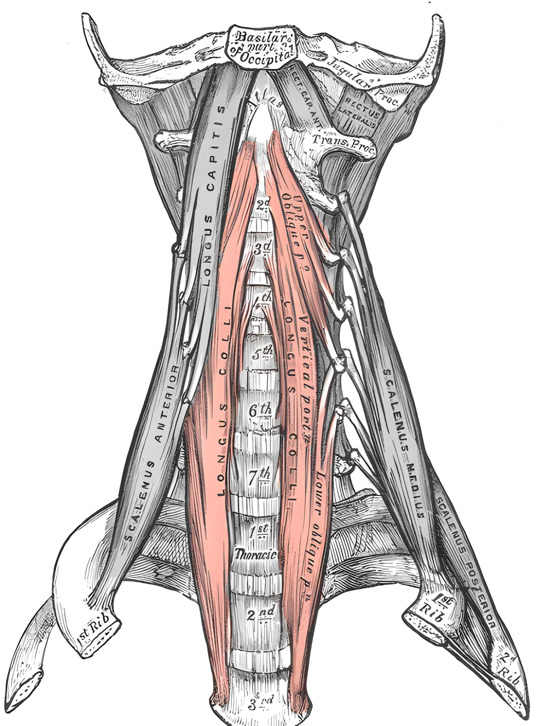
The paired Musculus longus colli is part of the prevertebral musculature.
Anatomy
The Musculus longus colli consists of three different fiber groups and therefore has an approximately triangular shape[^1].
Lateral superior fibers
The lateral superior fibers originate from the anterior tubercles of the transverse processes of the 5th through 2nd cervical vertebrae and insert on the anterior tubercle of the atlas.
Lateral inferior fibers
The lateral inferior fibers originate from the bodies of the 1st through 3rd thoracic vertebrae and extend to the anterior tubercle of the transverse process of the 6th cervical vertebra.
Medial fibers
The medial fibers extend from the bodies of the upper thoracic and lower cervical vertebrae to the bodies of the upper cervical vertebrae.
Function
Unilateral activation of the Musculus longus colli flexes and rotates the cervical spine to the side. Activation of both Musculi longi colli can flex the cervical spine forward[^1].
Innervation
The Musculus longus colli is innervated via the cervical plexus and Brachial plexus (C2 to C8) [^1]
Clinical relevance
During ACDF surgery, depending on the extent of the Musculus longus colli, it may be necessary to partially detach it at its medial attachment at the level being operated on.