A lumbar disc herniation is one of the most common neurosurgical conditions. It involves protrusion or sequestration of the intervertebral disc into the spinal canal or the neural foramen and can therefore cause severe pain and neurological deficits due to nerve root compression.
Localization
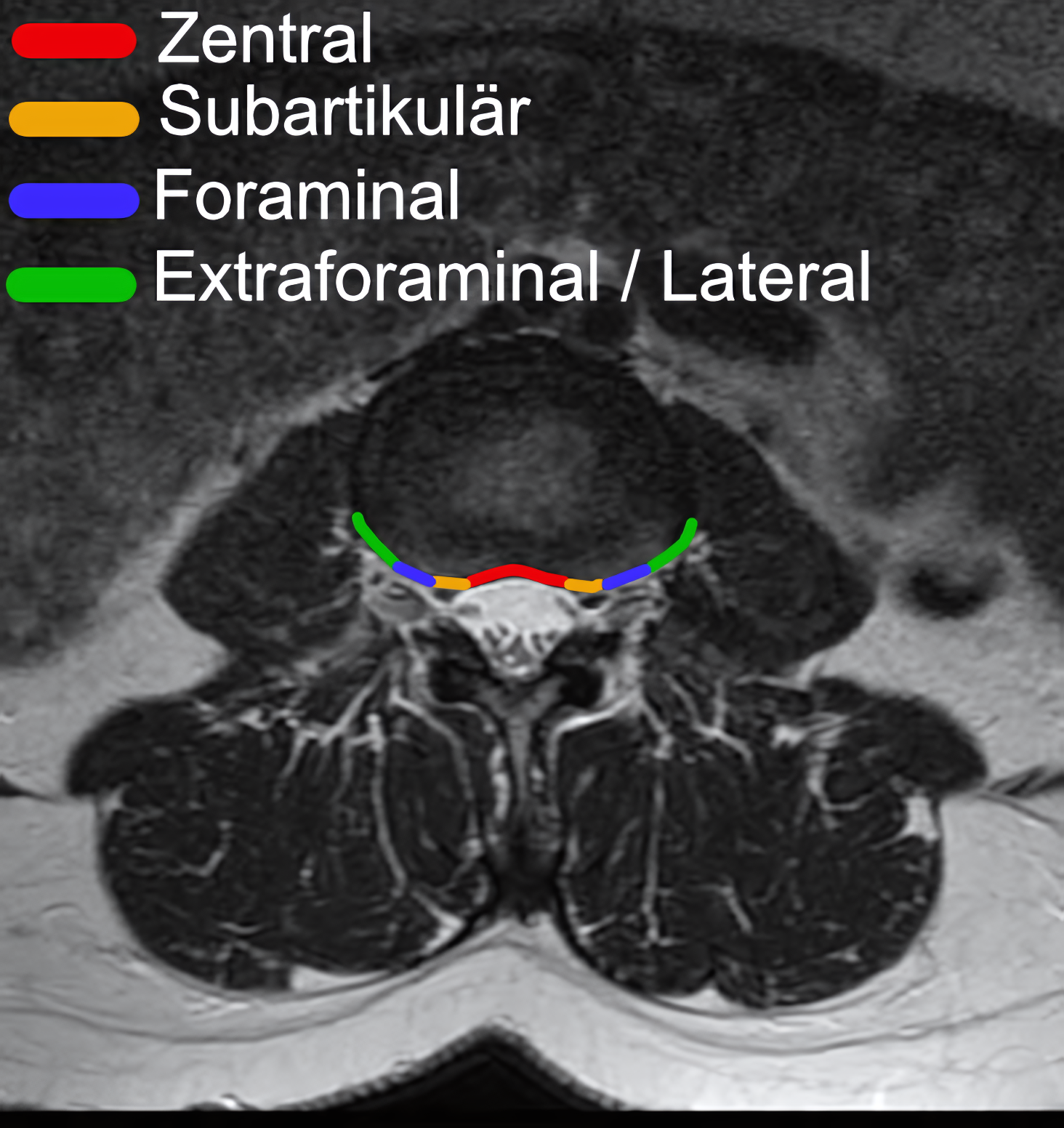
Depending on their location on axial imaging, lumbar disc herniations are classified as central, subarticular, foraminal, or extraforaminal disc herniations.
EnlargeNomenclature of lumbar disc herniations according to location, shown on an axial T2 MRI image.
Imaging
The gold standard for diagnosing lumbar disc herniations is MRI imaging.
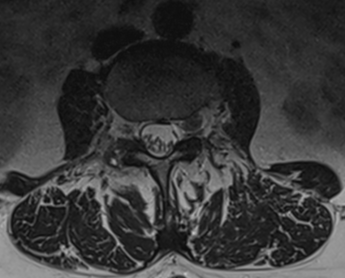
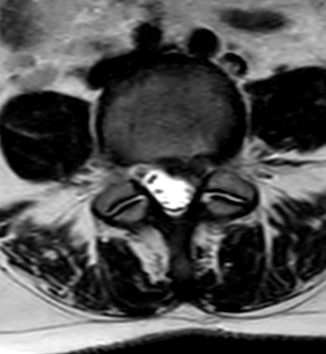
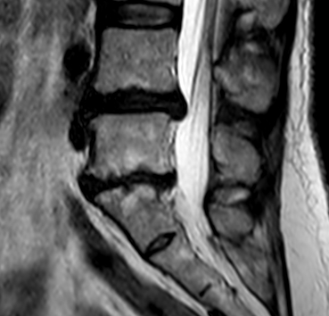
EnlargeLumbar disc herniation at the L5/S1 level on MRI.EnlargeAxial T2 MRI image of a patient with a left-sided intraforaminal disc herniation at L3/4.EnlargeAxial T2-weighted MRI image of a left-sided disc herniation at the L4/5 level.EnlargeSagittal T2-weighted MRI image of a patient with a lumbar disc herniation at the L4/5 level.
Figure
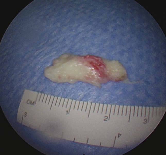
EnlargeAbbildung eines endoskopisch entfernten lumbalen Bandscheibenvorfalls.
Weekly update
Newsletter
Relevant neurosurgical studies, concisely summarized in a weekly email newsletter.
By subscribing, you consent to receiving the newsletter through Buttondown. Unsubscribe using the link in any email.
Privacy policy