Hemangioblastomas are vascular neoplasms of the central nervous system and occur predominantly in young adults. They are benign tumors located primarily in the Cerebellum and are classified as Grade 1 tumors according to the WHO classification.
WHO classification
According to the 2021 WHO classification, hemangioblastomas are classified as WHO Grade 1 tumors[^1].
Epidemiology
Hemangioblastomas are the most common primary brain tumors in the posterior cranial fossa in adults[^2]. Hemangioblastomas account for approximately 1.5 % to 2.5 % of all intracranial tumors and 7 - 8 % of all tumors of the posterior cranial fossa[^5].
Symptoms
The most common symptoms of hemangioblastomas of the posterior cranial fossa are attributable to increased intracranial pressure, followed by cerebellar symptoms[^5].
Imaging
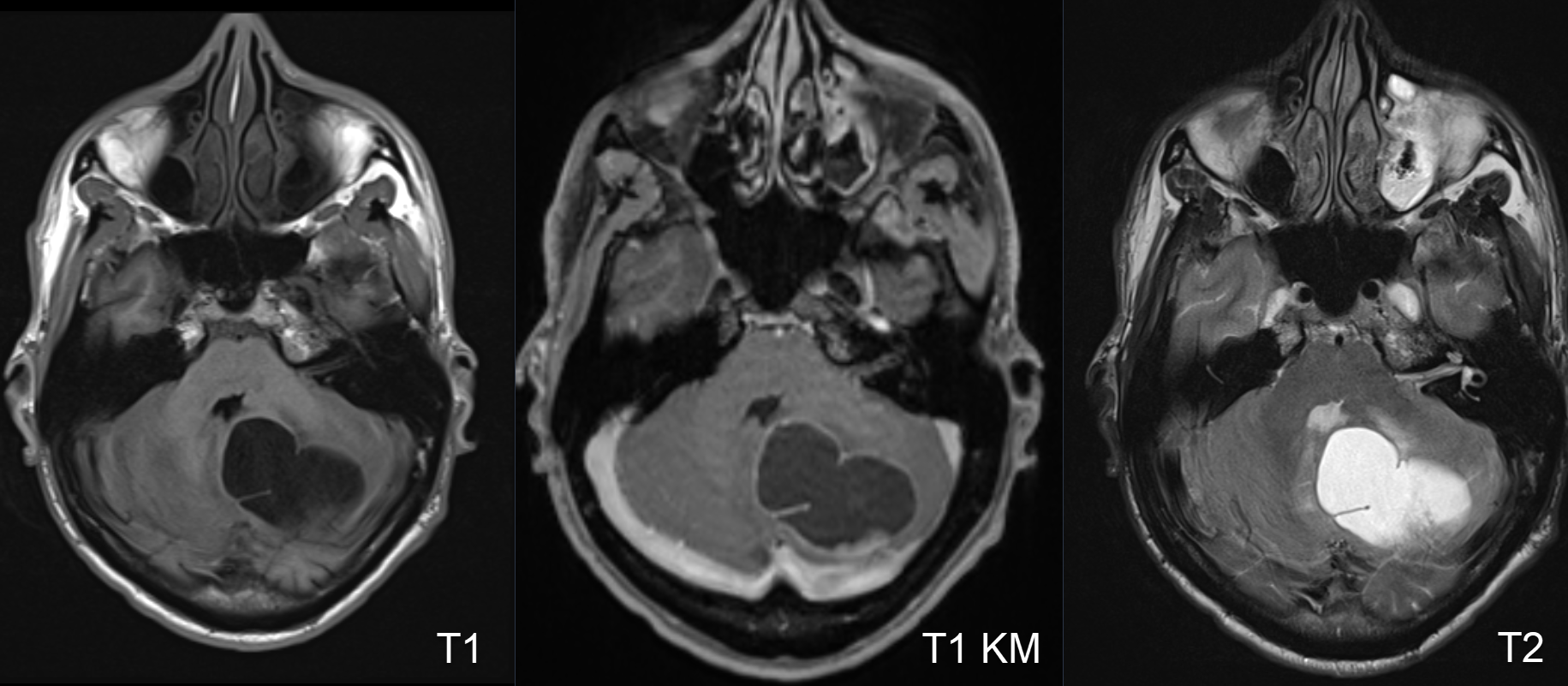
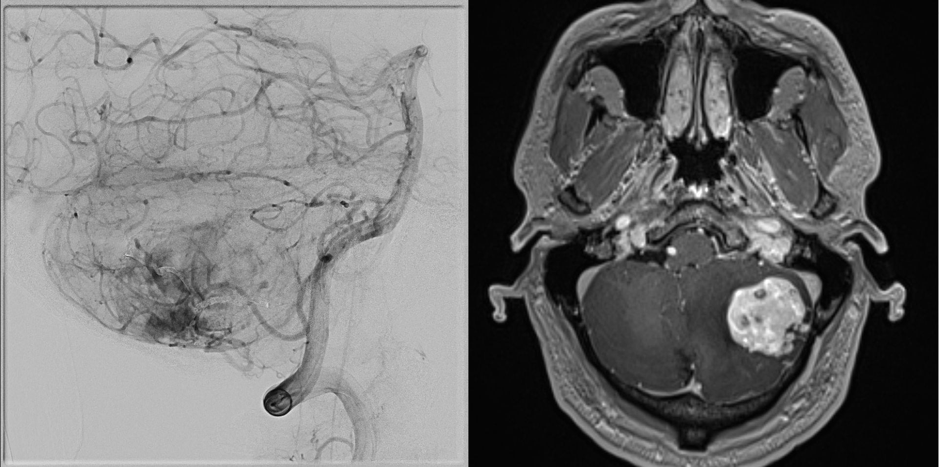
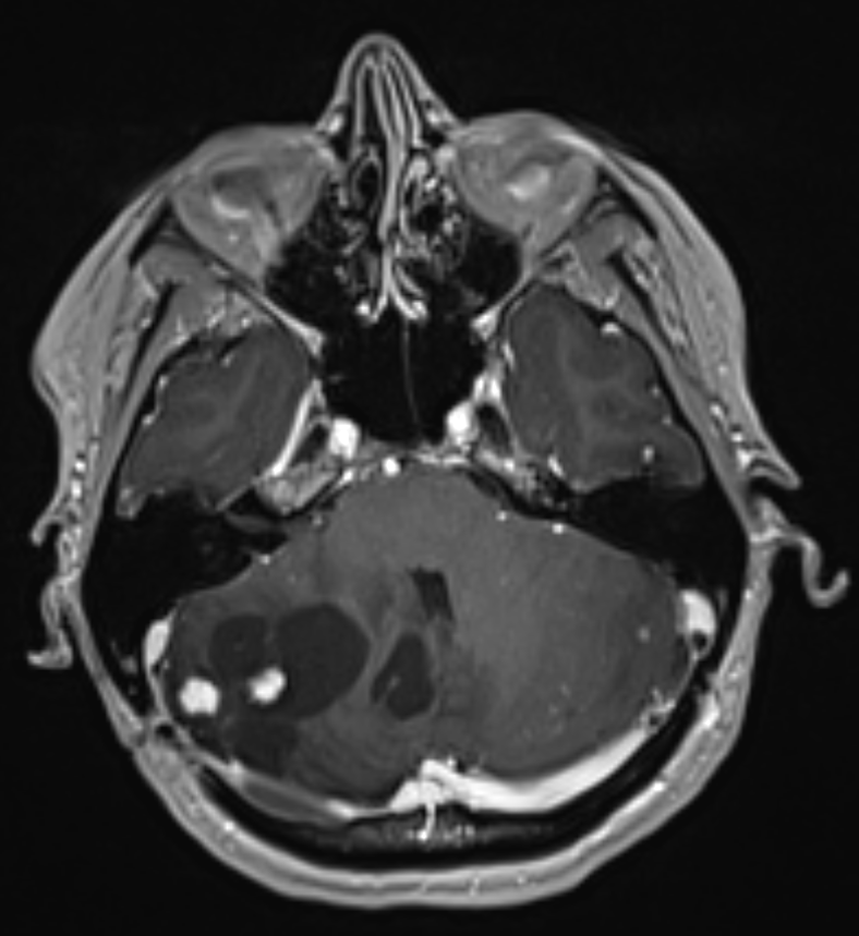
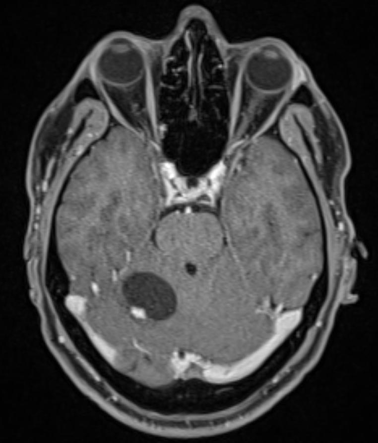
The gold standard for imaging hemangioblastomas is MRI imaging. digital subtraction angiography frequently demonstrates a highly vascularized nodule with feeding vessels, which is suggestive of a hemangioblastoma.
Associated diseases
In 25-40 % of cases, hemangioblastomas are diagnosed in patients with von Hippel-Lindau disease[^3]. In patients with von Hippel-Lindau disease, multiple hemangioblastomas may occur both cranially and spinally.
Pregnancy
In pregnant patients, there is a risk of rapid enlargement of the hemangioblastoma due to pregnancy[^4].