A craniopharyngioma is a benign tumor of the skull base that arises from epithelial remnants of Rathke’s pouch. Two distinct subtypes of craniopharyngioma are recognized.
Epidemiology
In the United States, the incidence of histologically confirmed craniopharyngioma is 0.16 / 100,000 inhabitants[^4].
Imaging
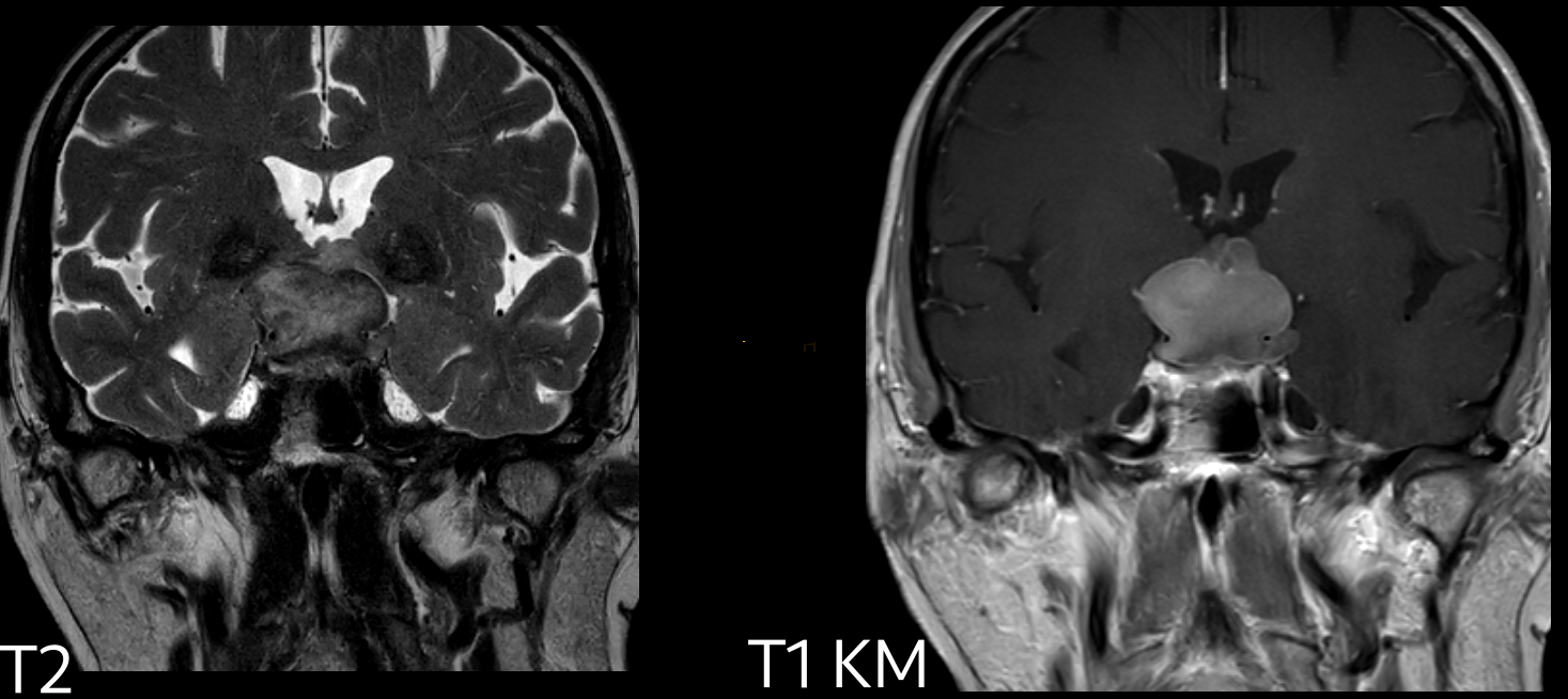
The imaging gold standard for evaluating craniopharyngiomas is MRI imaging. On MRI, craniopharyngiomas typically demonstrate heterogeneous contrast enhancement with solid and cystic components[^2]. On CT imaging, calcifications are seen in up to 80 % of cases of craniopharyngioma[^2].

Treatment
Surgical approach
The classic surgical approaches for craniopharyngiomas include the frontal interhemispheric, pterional, and endoscopic endonasal approaches[^2].
Subtypes
Adamantinomatous craniopharyngioma
Adamantinomatous craniopharyngioma is the most common type of craniopharyngioma. Adamantinomatous craniopharyngiomas are frequently associated with a beta-catenin (CTNNB1) mutation[^5].
Papillary craniopharyngioma
Papillary craniopharyngioma arises from metaplasia of adenohypophyseal cells[^1] and is less common than adamantinomatous craniopharyngioma. Papillary craniopharyngioma accounts for approximately 11-14 % of all craniopharyngiomas and occurs almost exclusively in adults[^3]. Papillary craniopharyngiomas are associated with a BRAF (V600E) mutation[^5]