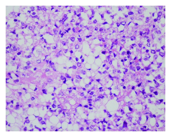
Cerebellar liponeurocytoma is an intrinsic brain tumor of the cerebellum (WHO grade 2).
Historical background
The first description of cerebellar liponeurocytoma was published in 1978 by Bechtel et al. [^3]. Cerebellar liponeurocytoma was first included in the 3rd edition of the WHO Classification of Tumors of the Central Nervous System in 2000 as a separate entity classified as a WHO grade 1 tumor. With improved recognition and longer follow-up, evidence of possible tumor recurrence accumulated. The recurrence rate was estimated at 60% during follow-up, with some cases even demonstrating aggressive behavior. Based on these findings, cerebellar liponeurocytoma was upgraded to WHO grade 2 in the 4th edition of the WHO Classification of Tumors of the Central Nervous System[^2].
Epidemiology
Cerebellar liponeurocytoma is a rare tumor and occurs slightly more frequently in women than in men (1.8:1)[^2].
Location
This type of tumor occurs exclusively in the posterior cranial fossa of adults[^1].
Imaging
On imaging, cerebellar liponeurocytomas tend to grow exophytically into the adjacent subarachnoid spaces. The fat attenuation on CT and T1 hyperintensity on MRI help distinguish this rare tumor from the more common Medulloblastomas or Ependymomas in adults.
CT
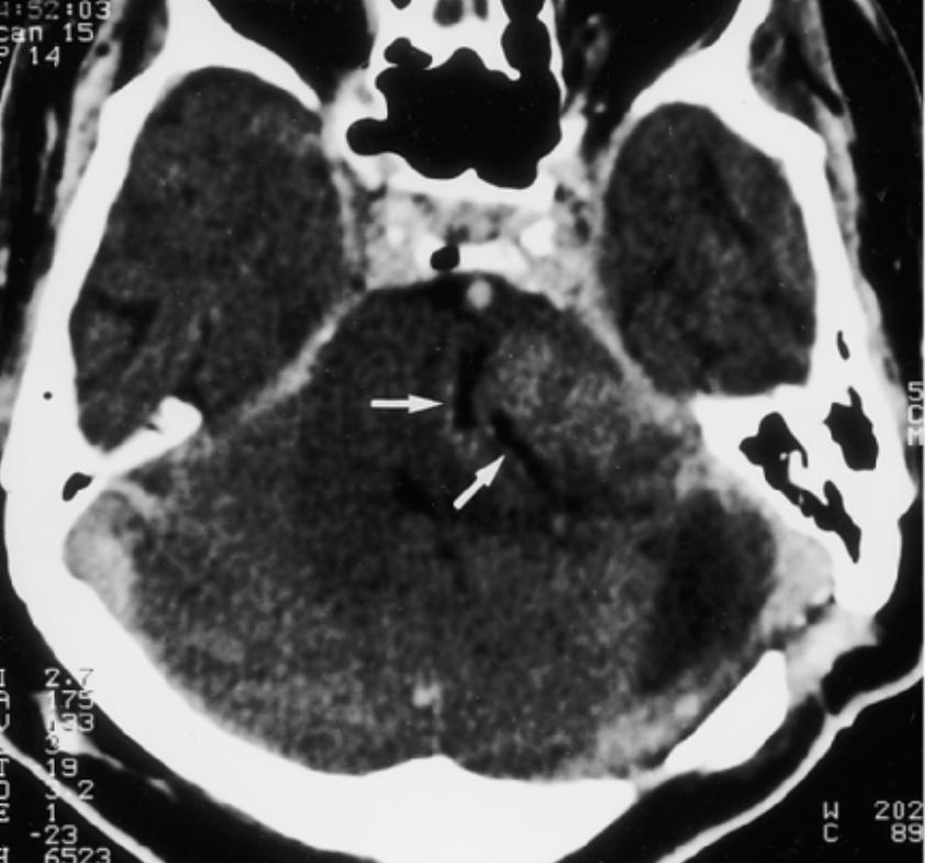
On CT imaging, the tumor typically appears as a hypodense mass with interspersed areas exhibiting the attenuation values of fat tissue[^4].
MRI
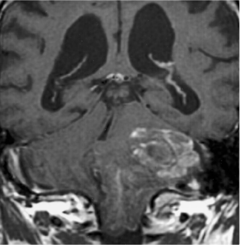
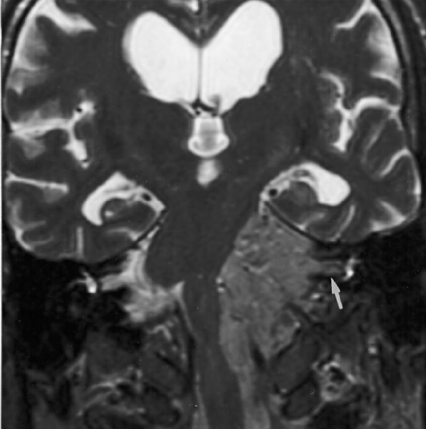
On T1-weighted MRI sequences, a cerebellar liponeurocytoma is hypointense with scattered foci of hyperintense signal and moderate contrast enhancement. On T2-weighted MRI sequences, the tumor is slightly hyperintense relative to the cortex, and edema is generally absent[^4].
Histopathology
These are neoplastic neurocytic cells that accumulate lipid.