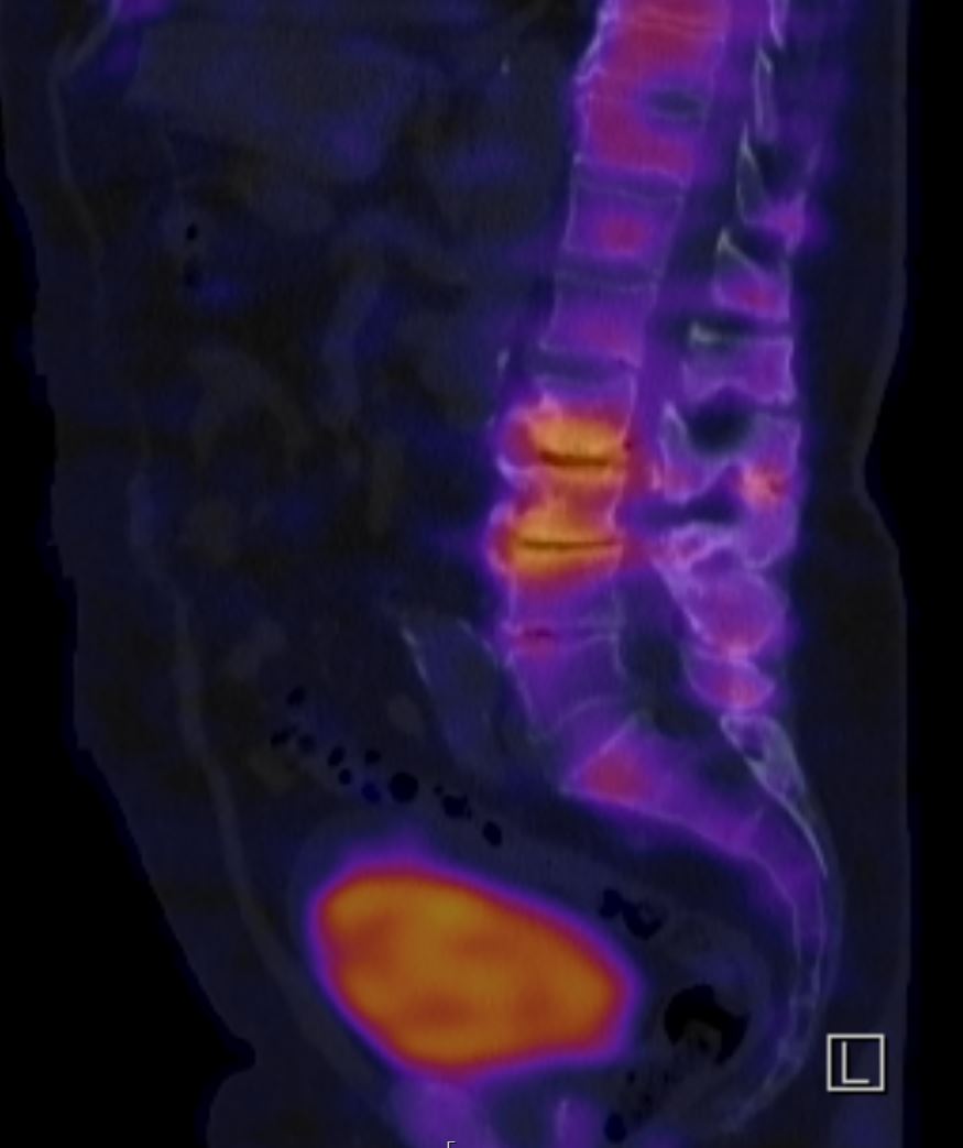
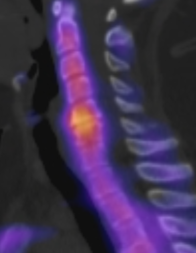
Single-photon emission computed tomography (SPECT)/CT is a hybrid imaging modality that plays an important diagnostic role, particularly in spinal neurosurgery. It combines the functional information provided by SPECT with the detailed anatomical images of the Computed tomography (CT). This fusion enables more precise localization and characterization of pathological processes in the Spine.
Principle of operation
During SPECT/CT, a small amount of a radioactive substance, known as a radiopharmaceutical, is injected into a vein. This accumulates in areas of increased bone metabolism, such as those occurring with inflammation, fractures, tumors, or osteoarthritic changes. A gamma camera detects the gamma radiation emitted by the radiopharmaceutical and uses it to create three-dimensional images (SPECT) showing metabolic activity in the bone. Immediately before or afterward, a CT examination of the same body region is performed. CT produces high-resolution cross-sectional images of the bony and soft-tissue structures. Through computer-assisted fusion of the SPECT and CT images, the “hot spots” (areas of increased metabolic activity) identified by SPECT can be precisely assigned to the anatomical structures shown on CT.