Schmorl Nodes

Last edit by Alaric Steinmetz on

Schmorl nodes are defined as herniation of intervertebral disc tissue into the vertebral body.

Imaging

Schmorl nodes can best be assessed on a sagittal MRI image. Symptomatic Schmorl nodes are usually acute and differ from chronic asymptomatic Schmorl nodes on MRI imaging[^1].

Classification of Schmorl nodes based on the MRI signal compared with the surrounding bone marrow[^2]:

Schmorl nodes

MRI <b>T1 sequence</b>

MRI <b>T2 sequence</b>

Symptomatic (Acute)

Hypointense

Hyperintense

Asymptomatic (Chronic)

Hyperintense

Hypointense

Schmorl's nodes MRI
Sagittal T2-weighted image of the lumbar spine showing Schmorl's nodes.
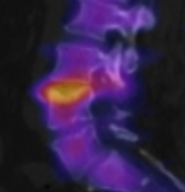
SPECT/CT of Schmorl node
Illustration of an activated Schmorl node on SPECT/CT of the lumbar spine.

Article information

Article created by
Last edited:
on
Revision history
Show revision history (2)

2 edits by 1 author

Edit · Alaric Steinmetz ·

No content change recorded

Article created on · Alaric Steinmetz

Author list:
Newsletter